By Prof. Somsanguan Asanyakhun
Head of LASIK Center Faculty of Medicine Chiang Mai University
What is Femtolasic?
Femto-LASIK is a laser refractive surgery (LASIK) that uses a femtosecond (FS) laser to cleave the cornea (Fig. 1) before using an excimer laser to shatter it. corneal texture to correct vision abnormalities as desired
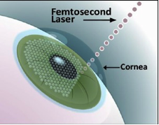
Figure 1. Femtosecond laser corneal separation.
Because femtolasik is a non-mechanical micokeratome LASIK surgery, which is a traditional corneal blade cleavage. It is called Blade-free LASIK, or Bladeless LASIK or all laser LASIK
How does the FS laser work?
FS laser is a laser that emits light energy at a wavelength of 1053 nm with a very short wavelength of 10 -15 seconds, which produces tiny shock waves and the formation of gas bubbles in the laser-treated tissue. When the air bubbles are completely absorbed by neighboring tissues Only cavitation bubbles are left (Fig. 2). The FS laser affects the tissue volume less than other lasers. The gas bubbles formed include carbon dioxide and nitrogen. will result in separation of tissue layers (photodisruption) without raising the temperature Therefore, it can be used in many types of refractive surgery.
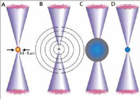
Fig. 2 shows the operation of the FS laser. (A) Absorption of multiphoton produces plasma. (B) Expands plasma and repels acoustic shock wave. (C) Cavitation bubble forms. (D) When contracted, gas bubbles are formed.
Why Femtolasik?
FS laser has been used since 2001 and has been gaining more and more popularity. Until now, corneal separating has been used for LASIK surgery instead of traditional corneal separating. microkeratomes Due to the advantages of corneal cleavage with FS laser compared to used Several traditional microkeratomes are as follows:
- Reduce the incidence of corneal separating complications such as corneal flap abnormalities such as corneal flap perforation. Buttonholes, free caps, irregular cuts, epithelial abrasions, etc. These factors can affect the value. clarity of vision after surgery
- Increase the accuracy and set the thickness of flap better
- More flap characteristics such as size and thickness can be selected. side cut angle, position and length of the hinge, etc.
- The flap can be cut thinner, 90-120 microns, while the traditional microkeratome can cut the flap as thin as 130-150 microns, thus making it possible for people with very impaired vision and thin corneas. who are unable to undergo LASIK surgery as well have traditional microkeratomes. It is possible to have LASIK surgery using the FS laser safely.
- Can reduce the chance of new astigmatism after LASIK (induced astigmatism) more.
- During corneal separating surgery If the suction loss is lost, the suction ring can be replaced immediately and the corneal layer can be separated to its original thickness, which the microkeratome cannot do.
- Less dry eye condition after surgery
- The angulation of the flap edge can be controlled, which influences the strength and wound healing. and prevent epithelium proliferation (epithelial ingrowth) better penetrate under the cornea lid
- After surgery, there is better contrast sensitivity, resulting in sharper images.
- The flap adhesion is stronger, thereby increasing the likelihood of flap dislocation after surgery. less from accidents
- The intraocular pressure is less therefore making it safer for the optic nerve terminals
- Less postoperative infection by separating the cornea microkeratome 2.4 times more likely to get infected than FS laser
Although FS laser corneal cleavage has the aforementioned advantages. But it has the following drawbacks.
- More likely to develop diffuse lamellar keratitis (DLK). DLK is inflammation of the cornea. which assumes It was caused by the accumulation of gas bubbles caused by the energy of the FS laser. It was found that inflammation was reduced when lasers were used at higher pulse rates because the overall energy was reduced (meaning that FS 60 kHz was better than FS 15 kHz and FS. 30 kHz), which has not been studied in FS 150 kHz and FS 200 kHz.
- The formation of the opaque bubble layer (OBL) is when gas bubbles accumulate in the corneal tissue or gas bubbles leak into the anterior chamber. This may interfere with the iris registration and pupil tracking systems during excimer laser ablation by OBL as a result of the gas bubbles generated by the FS laser being inadvertently absorbed by the surrounding tissue. thus inserted and accumulated between the corneal tissue layers OBL formation is associated with thick and small flap size and is common in the FS 15 kHz, the older FS laser. However, OBL or gas bubbles entering the anterior chamber It has no effect on long-term treatment results.
- Causes transient light-sensitivity syndrome (TLSS), a transient light sensitivity that occurs after surgery despite good visual acuity, presumably caused by inflammation, as symptoms improve with anti-inflammatory drugs. It is most common in older FS lasers such as the FS 6 kHz and FS 15 kHz, but is less common in the FS 30 kHz and FS 60 kHz.
How should I prepare? in Femtolasic surgery
In order to correct vision abnormalities with femtolasic achieved the desired results The following preparations should be made.
Preparation for eye assessment before surgery
1. If you have previously worn contact lenses, you must refrain from wearing them in order to return the cornea to its natural shape. By specifying the time to refrain from wearing contact lenses depending on the type of lens as follows
1.1 Soft contact lenses (soft contact lenses) that do not correct astigmatism, do not wear for 1 week.
1.2 Soft contact lenses To correct astigmatism (soft toric lens) stop wearing for 2 weeks
1.3 Rigid gas permeable lens, stop wearing for 1 month
2. On the day of the examination, sunglasses should be prepared. and someone took them back Because the iris must be dilated. To assess the condition inside the eye in detail, such as cataracts, glaucoma, macular degeneration, etc. Can't fight the light after dilation of the iris will take 3-6 hours, so it's not appropriate to drive back by yourself.
Preparing for Femto-Lasic Surgery on the Day of Surgery
1. Refrain from wearing the same contact lens that was prepared to assess the eye condition before surgery.
2. Refrain from using cosmetics on the face and around the eyes of all kinds
3. Refrain from using perfume, skin care products deodorant Scented hair oil or gel because it affects the operation of the laser
4. Wear comfortable, non-constrictive clothing, especially shirts. should be a wide neck or front cut with buttons to avoid scuffing against the eyes while removing because it may cause the flap to move
5. Able to eat normally
6. Will get a clean face. Especially around the eyelids and eyelashes. and inject antibiotics before entering the operating room
Practice during Femtolasik surgery
- After lying on the operating table (Fig. 3), the face is covered with sterile material. By opening only the eye area and receiving anesthetic drops in the eye that will be operated on Wait until the anesthetic takes effect.
- The bed will move to the FS laser to separate the corneal layer. An eye speculum will be inserted at this time. You will need to keep your eyes open, unstressed, and look into the aperture of the surgical camera. Then enter the docking process by inserting a suction ring to increase eye pressure. Once the eye pressure has reached the threshold, the doctor will insert a patient interface (PI) and adjust the cone to the surface of the cornea. Now the image will disappear.
- Once the flap's characteristics (e.g. location, canal length, position and hinge length) are determined, the FS laser is used to deliver energy to the cornea for separation. which if the FS 200 takes about 6 seconds
- After that, the bed will move to the excimer laser machine where the doctor will open the separated flap with the FS laser and use the excimer laser to grind the cornea to correct the vision as desired. With the EX 500, it takes about 1.4 seconds per 1 diopter, which means that with 5 diopter, it takes about 7 seconds.
The doctor will flush under the flap and put the flap back into place. And after waiting for the flap to attach to the eye for about 2-3 minutes, remove the speculum from the eye. Subsequently, an eye examination was performed after surgery. will allow him to rest for a while and be able to go home by covering the eye shield (eye shield) to prevent rubbing the eyes for 1 night
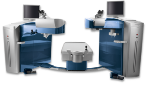
Figure 3 shows the FS laser FS 200 (left) and the Excimer laser EX 500 (right) of the WaveLight Refractive Suite (courtesy of LASIK Center, Faculty of Medicine. Chiang Mai University) Professor Somsanguan Asankhun, M.D.
How should I behave? After Femtolasic surgery
Proper behavior after femtolasic surgery It is also very important. Because it will help reduce or prevent complications after surgery. The following should be followed.
1. Cover the eyes should be closed before going to bed for 1 week to prevent rubbing the eyes unconsciously while sleeping, which may cause the flap to move.
2. You should be careful not to get water into your eyes when washing your face, bathing or washing your hair for 1-2 weeks to prevent infection under the flap, including refraining from swimming, sauna and scuba diving for 1 month.
3. Should refrain from makeup Especially eye makeup for 1-2 weeks to avoid makeup splashing into the eyes.
4. Wear sunglasses when outdoors. to avoid sunlight, wind and dust for about 1 month
5. Apply antibiotic and anti-inflammatory drops 4 times a day for 1-2 weeks together with artificial tears without preservatives. (non-preservative artificial tear) often following dry eye symptoms or as the physician deems appropriate
6. Come for a check-up by the doctor's appointment. or if abnormal Let's come to check immediately.
How should I check? After Femtolasic surgery
In general, the follow-up examination after femtolasic surgery Postoperative examinations will be done at least 6 times, 1 day, 1 week, 1 month, 3 months, 6 months, and 1 year after surgery to look at postoperative wounds, measure eyesight, and assess the condition inside the eye. Additional appointments may be made if abnormalities are found, such as inflammation, very dry eyes, or suspected infection, etc.
In case the patient is abroad or going to travel to study abroad Should be examined after surgery at least 2 times, 1 day and 1 week, and medical report results can be taken to be examined with an ophthalmologist abroad.
You can contact us for information at the LASIK Center, Faculty of Medicine. Chiang Mai University
Tel. 0-5393-9777 Call center 08-2766-6909 or www.facebook.com/cmulasik
Prof. Somsanguan Asanyakhun
Department of Ophthalmology Faculty of Medicine
Chiang Mai University