Nipt1

Noninvasive Prenatal Testing (NIPT)

Noninvasive Prenatal Testing (NIPT)

พญ.ลินลดา วิจักขณ์อุรุโรจน์
รศ.นพ.ชเนนทร์ วนาภิรักษ์


เซลล์ลูกในเลือดแม่ มี 2 ชนิดคือ Intact fetal cell และ cell- free fetal DNA(1)

  1. Intact fetal cell คือเซลล์ลูกที่พบในเลือดแม่และคงอยู่ในกระแสเลือดแม่ได้ถึง 10ปี หลังคลอด เป็นเซลล์ที่พบได้ในปริมาณน้อย และเป็นส่วนหนึ่งในการก่อให้เกิดโรค autoimmune diseaseในแม่ เช่น SLE ,scleroderma, Hashimoto Thyroiditis เป็นต้นในทางปฏิบัติไม่สามารถนำมาใช้ในการ prenatal diagnosis ได้เนื่องจากเป็นเซลล์ที่มีปริมาณน้อย คงอยู่นานหลังคลอด และแยกจากเลือดแม่ได้ยาก
  2. Cell-free fetal DNA คือ สิ่งที่ปล่อยจาก apoptotic placental trophoblast เข้าสู่เลือดแม่ สามารถตรวจพบได้ตั้งแต่ อายุครรภ์ 7 สัปดาห์ และพบในกระแสเลือดแม่ถึง 3-6% และปริมาณจะเพิ่มขึ้นตามอายุครรภ์ และขับออกจากกระแสเลือดแม่ภายใน1-2 ชั่วโมงหลังคลอด โดยเซลล์ชนิดนี้เป็นสิ่งที่สามารถนำมาใช้ตรวจหา โรคทางพันธุกรรม( single gene disorder), หมู่เลือดในลูก( fetal Rh D genotype evaluation), เพศลูก(Fetal Sex determination) และ การตรวจคัดกรอง aneuploidy(NIPT)

NIPT ( Noninvasive prenatal testing )


เป็นวิธีหนึ่งในการตรวจคัดกรองเพื่อหาfetal aneuploidy โดยใช้ cell- free fetal DNA มา sequence DNA fragment เพื่อดูอัตราส่วนของ โครโมโซมที่สงสัยว่ามีปริมาณมากเกินปกติหรือไม่ เช่น หากทารกมีภาวะเป็น Trisomy 21 จะตรวจพบDNA sequence ของ โครโมโซมคู่ที่ 21 มากกว่าปกติ

วิธีนี้มี ความไวในการตรวจคัดกรอง(sensitivity) 98% และอัตราการเกิดผลบวกลวง(false positive rate)<0.5% ในการตรวจคัดกรองโรคกลุ่มดาวน์(Down’s syndrome)ซึ่งจัดว่าเป็นวิธีที่มีdetection rate ที่สูงกว่าวิธีตรวจคัดกรองอื่นๆตามตารางที่1(1)

{tabulizer:style[gr.alterora.elemental_1_green.css]}

Screening Strategy Detection Rate(%)
First trimester screening (NT+PAPP-A+hCG) 79-87
NT alone 64-70
Triple test 61-70
Quadruple Test 74-81
Integrated screening 94-96
Stepwise sequential screen 90-95
Contingent sequential screen 88-94
Cell-free fetal DNA 98

ตามความเห็นจาก ACOG (American College of Obstetrician and Gynecologists) และ sMFM opinion ปี 2012 แนะนำว่าNIPT เป็นวิธีการตรวจคัดกรองที่มีประสิทธิภาพที่สุดในกลุ่ม high risk pregnancy ซึ่งข้อบ่งชี้ที่สามารถแนะนำให้ตรวจคัดกรองได้แก่(2)

  1. สตรีตั้งครรภ์ที่มีอายุ ≥ 35 ปี
  2. มีหลักฐานจาก ultrasound ตรวจพบว่าทารกมีความเสี่ยงสูงต่อการเกิด aneuploidy
  3. มีประวัติบุตรคนก่อนเป็น fetal aneuploidy
  4. เคยตรวจคัดกรองด้วยวิธีอื่น เช่น serum marker แล้วพบว่าทารกมีความเสี่ยงสูงต่อการเกิด fetal aneuploidy
  5. สตรีตั้งครรภ์หรือสามีตรวจพบเป็น balanced Robertsonian translocation ของโครโมโซมคู่ที่21หรือ13

Consensus statement จาก ISUOG (International Society of Ultrasound in Obstetrics and Gynecology) ได้กล่าวถึงประเด็นเพิ่มเติมดังนี้(3)

  1. หาก ทำ ultrasound anomaly screening แล้วพบว่ามี structural anomaly แนะนำให้ทำ fetal karyotyping แม้ว่าจะตรวจพบว่า ผล NIPT ปกติก็ตาม
  2. สตรีตั้งครรภ์ทุกรายควรได้รับการทำ first-trimester ultrasound แม้ว่าจะทำ NIPT หรือไม่ก็ตาม
  3.  เสนอ 3 ทางเลือกเพื่อการประเมิน fetal trisomy 13,18,21 ได้แก่
    • Maternal age + NT+/-serum marker
    • PND in high risk group
    • NIPT เป็น primary screening แต่อย่างไรก็ตาม การใช้NIPT ใน low risk group ยังไม่มีข้อมูลการศึกษาที่เพียงพอ อีกทั้งประชากรกลุ่มนี้เป็นกลุ่มที่มี PPV ต่ำอยู่แล้ว ปัจจุบันจึงแนะนำให้ใช้เฉพาะในกลุ่ม intermediate group
  4. หากทำ NIPT แล้วพบว่าผลปกติ จะไม่มีความจำเป็นที่จะต้องทำ genetic sonogram (soft marker ของ Down’s syndrome ) เนื่องจาก มี false positive rate ที่สูงกว่าและมี PPV ที่น้อยกว่าเมื่อเทียบกับ NIPT
  5. ไม่แนะนำให้ใช้ NIPT แทนการทำ PND ในกลุ่ม high risk group จากการทำ serum marker screening
  6. หากผลตรวจ NIPT ปกติ ไม่มีความจำเป็นต้องตรวจ NTและ serum marker ซ้ำอีก
  7. ความแม่นยำในการใช้ NIPT ในกรณีที่เป็นครรภ์แฝด ยังไม่เป็นที่สรุปแน่ชัด

Bioinformatics techniques in NIPT(4-9)

ตั้งแต่ ค.ศ.1999 มีการศึกษาพบว่าสามารถสตรีตั้งครรภ์ที่ทารกในครรภ์เป็น Down’s syndrome จะมีปริมาณ fetal DNA ในเลือดแม่ในระดับที่สูงกว่าครรภ์ปกติ จึงมีการใช้เทคนิคต่างๆในการตรวจหาปริมาณ DNA ดังกล่าวได้แก่ DNA Methylation, circulating fetal RNA analysis ,allelic ratio measurement อย่างไรก็ตามวิธีดังกล่าวต้องใช้ markerที่หลากหลายในการตรวจเนื่องจากไม่สามารถหา markerที่เป็นตัวแทนของทุกtest ได้ จึงทำให้การตรวจมีข้อจำกัดมาก(4)

ในปี ค.ศ.2007 ได้มีการศึกษาวิธีการตรวจโดยใช้ digital PCR techniqueซึ่งเป็นการนับปริมาณ DNA moleculeและหากยิ่งมีปริมาณมาก ประสิทธิภาพในการแยก DNA ก็จะสูงขึ้นตามลำดับดังนั้นการใช้วิธีนี้จะมีข้อจำกัดในรายที่มีปริมาณของ cffDNAน้อย(4, 8, 9)

ในปี ค.ศ.2008 มีการศึกษาเกี่ยวกับการนำ MPS(Massively parallel sequencing)มาใช้ในการหาปริมาณDNA ซึ่งพบว่ามีข้อดีกว่าการใช้ digital PCRเนื่องจาก digital PCR จะตรวจพบเฉพาะ DNA ที่จำเพาะต่อ RNA primer เท่านั้น ในปัจจุบัน MPSที่ใช้อย่างแพร่หลายมี 2 วิธีคือ 1.Shotgun sequencing 2.Targeted sequencing(4)

1. Shotgun sequencing(5)

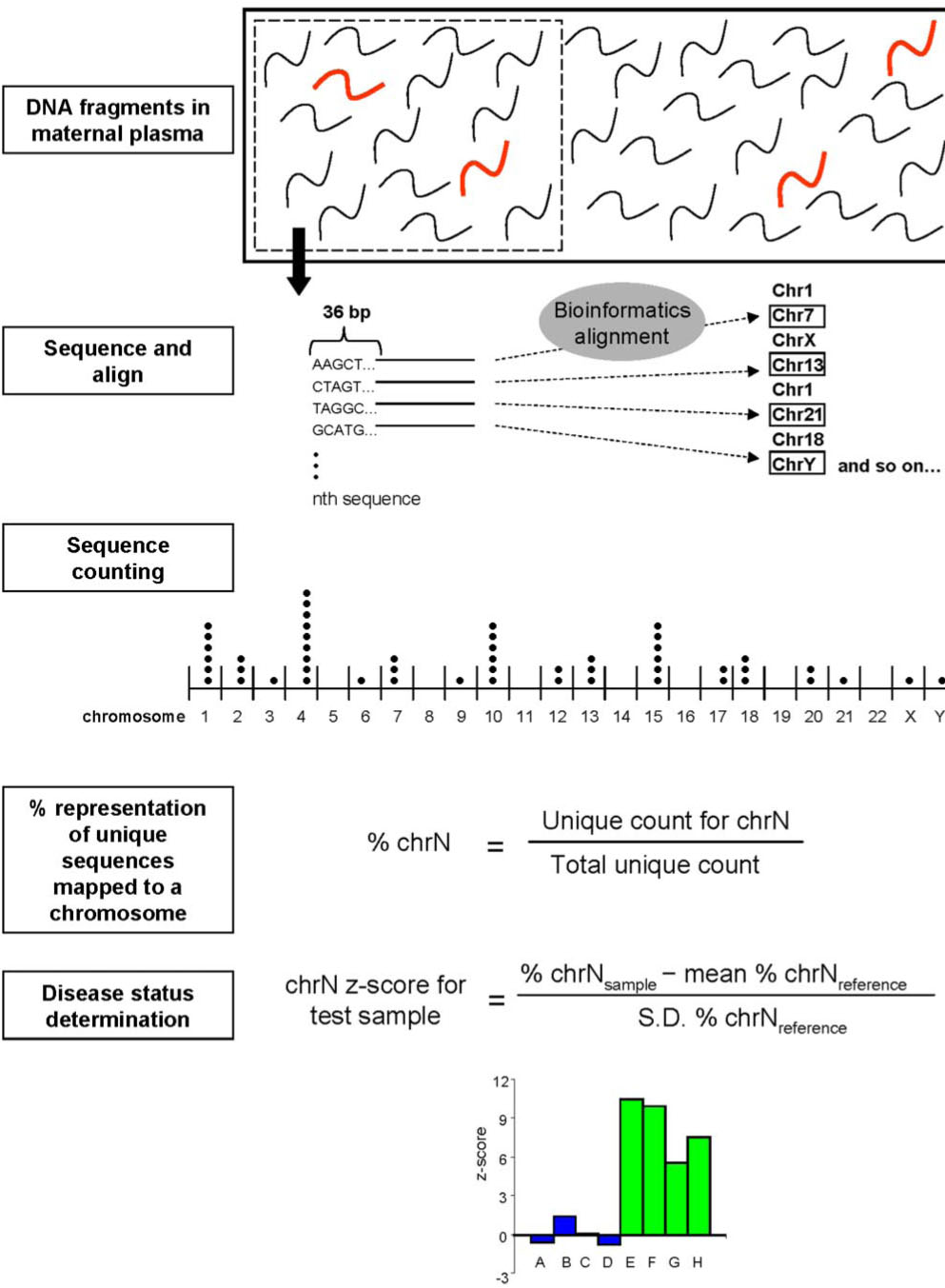
หลักการ : DNA ใน maternal serum จะถูกนำมาsequence แบบสุ่มในรูปแบบอัตราส่วนของ DNA ของโครโมโซมที่เราสนใจกับ DNAของโครโมโซมอื่นใน genomeดังแสดงตามรูปที่1

ข้อดีคือ ขั้นตอนในการ DNA sequencingของโครโมโซมที่เราสนใจจะไม่ขึ้นกับ genomic location ทำให้สามารถตรวจพบ genomic aberration ทุกที่ใน genomeและสามารถหา fetal whole genome sequencing จาก maternal serum ได้

Nipt1

2. Targeted sequencing(6, 7, 10)

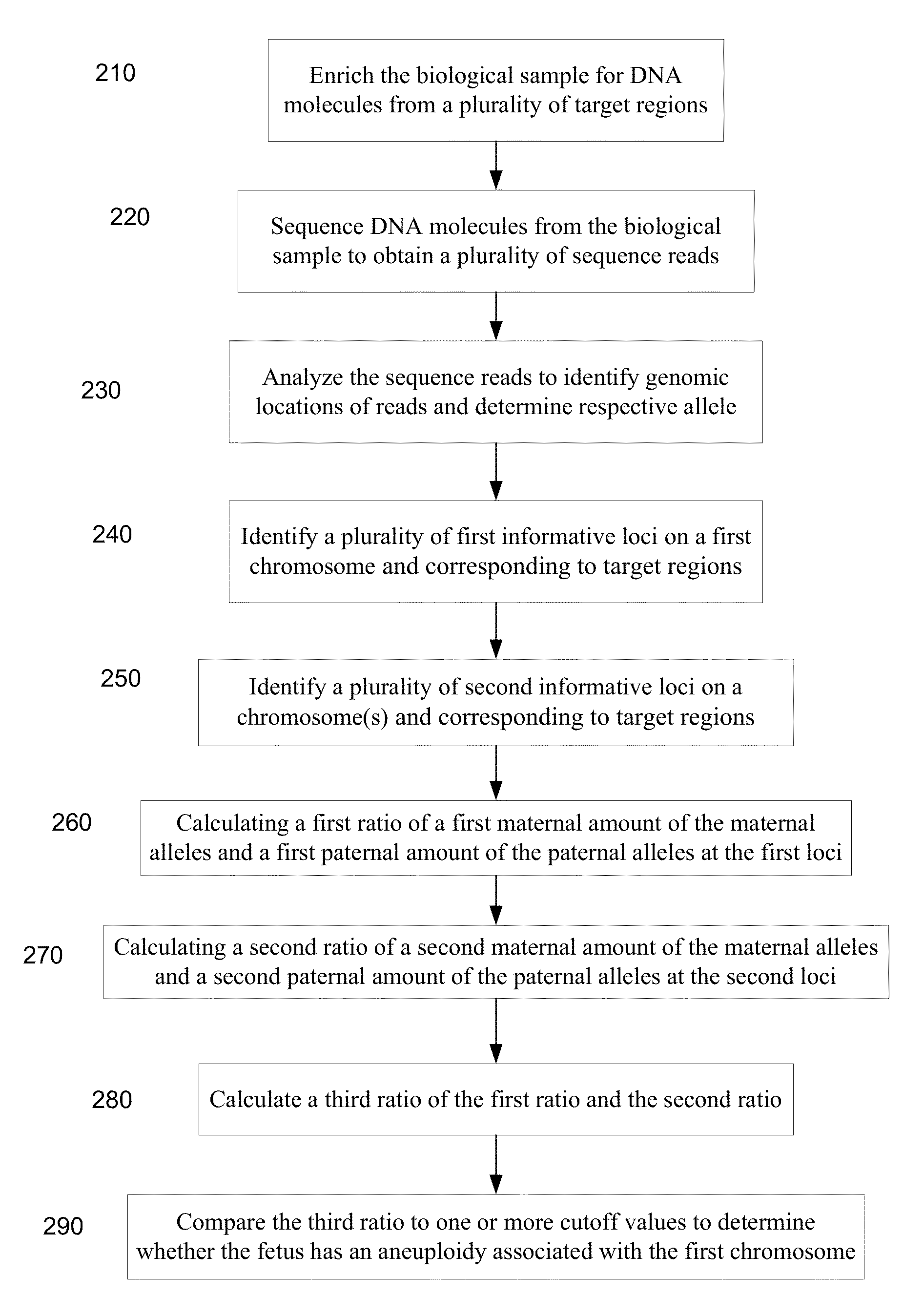
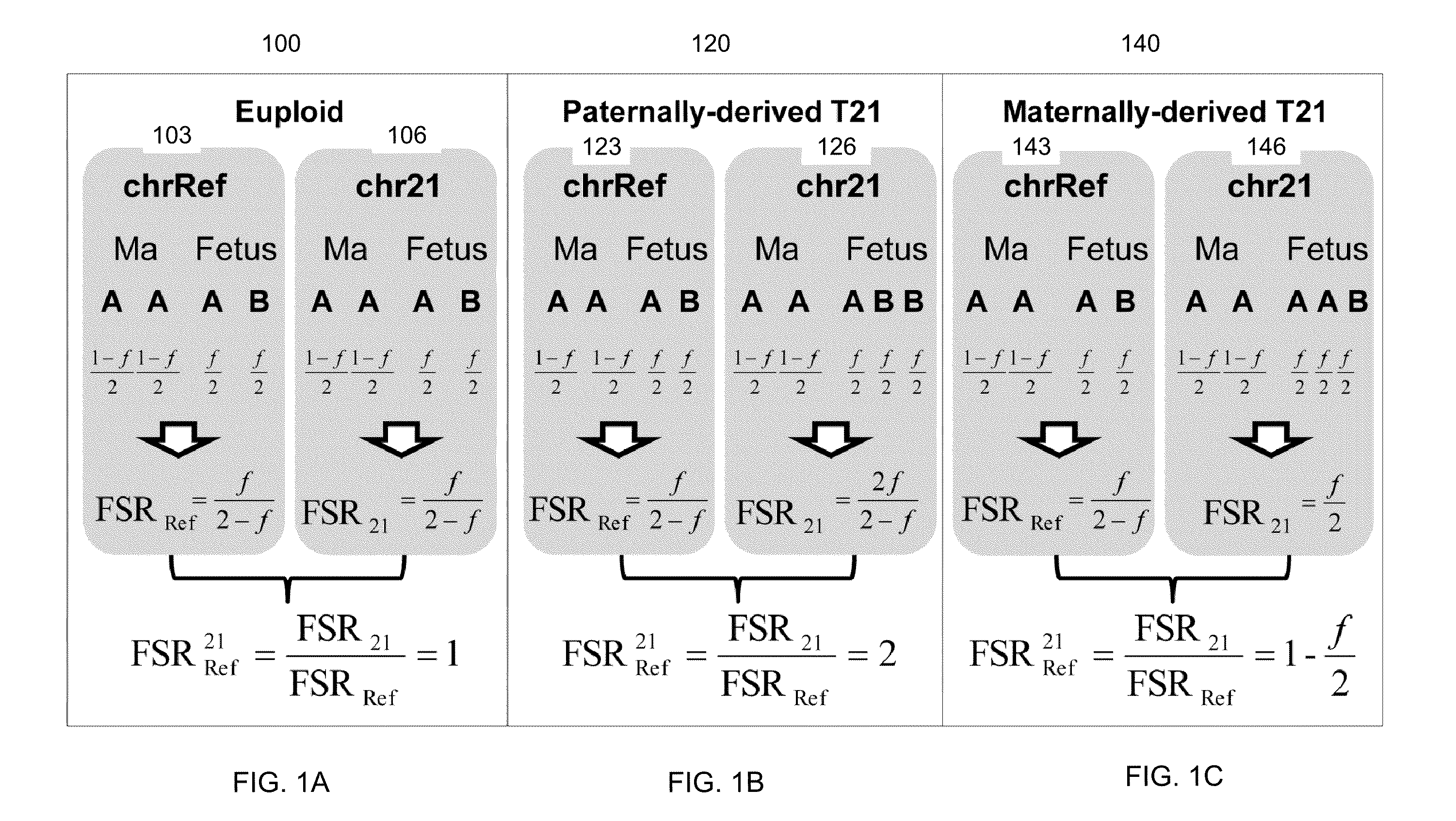
หลักการ:เป็นการเลือกsequenceเฉพาะ regionของgenomeที่มีโครโมโซมที่เราสนใจ(targeted region) เทียบกับ regionอ้างอิง ในปัจจุบัน มี 2วิธีที่ใช้คือ 1.ใช้ Digital Analysis of Selected Region essay (DANSR)(10)กับ targeted genome ที่ไม่มีความแตกต่างระหว่างบุคคล 2.ใช้วิธี allelic ratio analysis หากtargeted genome มี polymorphic ในกลุ่มประชากร ตามแสดงในรูปที่ 2

ข้อดี คือ sequencing power สูงและสามารถเลือก region ที่ต้องการให้ sequence ได้ แต่ข้อเสียคือ ต้องมี panel ที่จำเพาะต่อการทำ DNA hybridization probe, PCR primer

Nipt2

Nipt3

Possible causes of errors

RCOG( Royal College of Obstetricians and Gynaecologists )ได้กล่าวถึงข้อควรระวังในการใช้ NIPT ในกรณีต่างๆต่อไปนี้(4)

1. Early Gestational age

ปริมาณของ cffDNAเพิ่มขึ้นตามอายุครรภ์ ดังนั้นหากตรวจในช่วง early gestation อาจพบปัญหา false negative ของ Y chromosome และ RhDในขณะที่ aneuploidy screening จะสามารถตรวจพบความผิดปกติได้เมื่อมีปริมาณ fetal cell อย่างน้อย 4-5% หรือตั้งแต่หลัง GA 10 wkเป็นต้นไป ดังนั้นหากคนไข้ต้องการตรวจ NIPT ควรทำ ultrasound เพื่อยืนยันอายุครรภ์ก่อนทุกครั้ง

2. Maternal Obesity

จากการศึกษาพบว่าในมารดาที่น้ำหนักมากจะพบปริมาณของ cffDNAได้น้อยกว่าคนที่น้ำหนักปกติ โดยกลไกยังไม่เป็นที่ทราบแน่ชัดแต่เชื่อว่าเกิดจากการที่มีปริมาณของ maternal plasma DNA ที่เพิ่มขึ้น หรืออาจเกิดจาก blood volume ที่มากขึ้นและเกิด hemodilutionจึงทำให้ปริมาณ cff DNA ลดลงอย่างมีนัยสำคัญ จึงเป็นข้อที่ควรพิจารณาและแจ้งให้คนไข้ทราบก่อนทุกครั้ง

3. Multiple pregnancies

ในการตั้งครรภ์แฝดชนิด Monochorionการตรวจจะให้ผลที่มีประสิทธิภาพมากกว่า Singleton pregnancy เนื่องจากหากจะพบปริมาณ cff DNA ได้เป็น 2 เท่าของครรภ์ปกติ แต่ในรายที่เป็นครรภ์แฝดชนิด Dichorionการแปลผลจะทำได้ยากกว่าเนื่องจากอาจมีแฝดปกติและผิดปกติในครรภ์เดียวกันเช่นเดียวกับtwin pregnancy with one vanishing twin อย่างไรก็ตามมี 2การศึกษาที่รายงานว่าการใช้วิธี shotgun-MPS สามารถแปลผลในรายที่เป็น twin pregnancy ได้และยังสามารถบอก zygosityของ twinได้อีกด้วย

4. Placental mosaicism

เนื่องจาก cffDNAมาจาก apoptotic trophoblast ดังนั้นหากมีภาวะ CPM (Confined Placental Mosaicism )ซึ่งพบได้1% แต่หากมีภาวะนี้ จะส่งผลให้เกิดการแปลผลโครโมโซมผิดพลาดเกิดเป็น false positiveได้ทั้งที่ทารกปกติ ดังนั้นจึงต้องมีการทำ confirmatory test ด้วย PNDทุกครั้งหากผล NIPT ผิดปกติ

5. Maternal condition

หากมารดามีภาวะ mosaicism หรือมีmalignancy condition อยู่ จะทำให้เกิดfalse- positive ได้

Comparison between commercial NIPT

ในปัจจุบันได้มีการใช้ NIPT ในการ screening โดย commercial kit จากบริษัทที่มีในปัจจุบันได้แก่ NIFTY, NIPS, Harmony และ Panorama โดยมีคุณสมบัติแตกต่างกันเล็กน้อยดังตาราง

{tabulizer:style[gr.alterora.elemental_1_blue_green.css]}

NIFTY NIPS Harmony Panorama
Chromosome coverage All All 13,18,21,X,Y 13,18,21,X,Y
Timing to Test GA10 wk (singleton) / GA12wk (Twin) GA10wk (both) GA10wk (both) GA9 wk
Available for twin Yes Yes(Monozygote) Yes No
Available for triplet Yes No No No
Surrogate mother Yes Yes Yes No
Insurance for false positive rate 1,000,000 baht 2,000,000baht No No
Resample 1-3% 1-3% 1-3% 5-12%
Time to report 10 days 8-12days 8-12days 10-14days

บทสรุป

  1. สตรีตั้งครรภ์ทุกรายควรได้รับคำแนะนำในการตรวจคัดกรองต่อความเสี่ยงในการมีลูกเป็นtrisomy
  2. มีการคัดกรองมีหลายรูปแบบ ควรเลือกตามความพร้อมของทรัพยากรและบุคคล
  3. NIPT เป็นวิธีการตรวจคัดกรองที่ได้รับการยอมรับว่ามี ความไวสูงสุดและผลบวกลวงน้อยที่สุด
  4. แม้ว่า NIPT จะเป็นวิธีการคัดกรองที่ได้ผลดีแต่ก็ไม่สามารถนำมาใช้แทนการตรวจวินิจฉัยได้
  5. เนื่องจาก NIPT คือการตรวจหาปริมาณ cell free fetal DNA(cffDNA) ดังนั้นยิ่งอายุครรภ์มากก็จะตรวจพบได้ในปริมาณที่มากขึ้น ปกติแล้วจะแนะนำให้ตรวจได้ตั้งแต่อายุครรภ์ 10 สัปดาห์เป็นต้นไป
  6. นอกจากประโยชน์ในการตรวจคัดกรองเรื่อง fetal aneuploidy แล้ว NIPT ยังสามารถใช้ในการตรวจหา RhD blood group,Single gene disorder, fetal sex determination ได้อีกด้วย

เอกสารอ้างอิง

  1. F.Gary Cunningham KJL, Steven L.Bloom,Catherine Y.Spong,Jodi S.Dashe,Barbara L. Hoffman,Brian M.Casey,Jeanne S. Sheffield. Williams obstetrics. 2014 ed. New York: McGraw-Hill Education; 2014.
  2. Committee Opinion No. 545: Noninvasive Prenatal Testing for Fetal Aneuploidy. Obstetrics & Gynecology. 2012;120(6):1532-4 10.097/01.AOG.0000423819.85283.f4.
  3. Salomon LJ, Alfirevic Z, Audibert F, Kagan KO, Paladini D, Yeo G, et al. ISUOG consensus statement on the impact of non-invasive prenatal testing (NIPT) on prenatal ultrasound practice. Ultrasound in Obstetrics & Gynecology. 2014;44(1):122-3.
  4. Scientific Impact Paper No. 15: Non-invasive Prenatal Testing for Chromosomal Abnormality using Maternal Plasma DNA. The Obstetrician & Gynaecologist. 2014;16(2):148-.
  5. Chiu RWK, Chan KCA, Gao Y, Lau VYM, Zheng W, Leung TY, et al. Noninvasive prenatal diagnosis of fetal chromosomal aneuploidy by massively parallel genomic sequencing of DNA in maternal plasma. Proceedings of the National Academy of Sciences of the United States of America. 2008;105(51):20458-63. PubMed PMID: PMC2600580.
  6. Liao GJW, Chan KCA, Jiang P, Sun H, Leung TY, Chiu RWK, et al. Noninvasive Prenatal Diagnosis of Fetal Trisomy 21 by Allelic Ratio Analysis Using Targeted Massively Parallel Sequencing of Maternal Plasma DNA. PLoS ONE. 2012;7(5):e38154. PubMed PMID: PMC3362548.
  7. Zimmermann B, Hill M, Gemelos G, Demko Z, Banjevic M, Baner J, et al. Noninvasive prenatal aneuploidy testing of chromosomes 13, 18, 21, X, and Y, using targeted sequencing of polymorphic loci. Prenatal Diagnosis. 2012;32(13):1233-41.
  8. Lo YMD, Lun FMF, Chan KCA, Tsui NBY, Chong KC, Lau TK, et al. Digital PCR for the molecular detection of fetal chromosomal aneuploidy. Proceedings of the National Academy of Sciences of the United States of America. 2007;104(32):13116-21. PubMed PMID: PMC1934923.
  9. Fan HC, Quake SR. Detection of Aneuploidy with Digital Polymerase Chain Reaction. Analytical Chemistry. 2007 2007/10/01;79(19):7576-9.
  10. Sparks AB, Wang ET, Struble CA, Barrett W, Stokowski R, McBride C, et al. Selective analysis of cell-free DNA in maternal blood for evaluation of fetal trisomy. Prenatal Diagnosis. 2012;32(1):3-9.
Read More

Pelvic anatomy and clinical correlation

Pelvic anatomy and clinical correlation

พญ.ลินลดา วิจักขณ์อุรุโรจน์
รศ.นพ.กิตติภัต เจริญขวัญ


1. Anatomy of Skin and Abdominal Wall

Boundary

  • Superior border : rib 7-12
  • Inferior border : iliac crest,inguinal ligament, pubic bone
  • Posterolateral : lumbar spine and adjacent muscle

Skin Line

1. Langer Line

เป็นเส้นที่ดึงขอบของผิวหนังให้ห่างจากกกันเวลาลงมีดผ่าตัด ใน lower abdomen จะเป็นในแนว horizontal ดังนั้น การลงแผลแนวขนานจะเกิดแผลเป็นน้อยกว่าการลงแผลแนวดิ่ง

2. Arcuate Line

เป็นจุดบรรจบระหว่าง posterior aponeurosis ของ Internal oblique muscle กับ aponeurosis ของ Transversalis muscle ที่ต่ำกว่าเส้นนี้ลงไป ลำไส้จะแยกกับ abdominal wall ด้วย peritoneum และ oponeurosisของ intenal oblique muscle จะอยู่เหนือ rectus muscle

3. Linea Nigra and Linea Alba

  • Linea Alba : เป็นinsertion ของแนว Fascia ของ External Oblique, Internal Oblique และ Transversalis muscle
  • Linea Nigra : เป็น hyperpigment line ที่skin ตามแนวของ linea alba

Fascia

1.Superficial Fascia

1.1 Camper’s fascia อยู่ชั้นบนสุดมี fat มากแต่ fibrous tissue น้อย และต่อเนื่องไปเป็นชั้น superficial fatty layer ของ perineum

1.2 Scarpa’s fasciaเป็นชั้นที่อยู่เหนือ rectus sheath เป็นชั้นที่มี fibrous tissue มากเมื่อเทียบกับ fat และเป็นชั้นที่ต่อเนื่องไปเป็น Colles fascia (Superficial perineal fascia ) และ fascia Lataที่ขา

2. Rectus Sheath

จุดที่อยู่เหนือ Arcuate Lineจะมี anterior and posterior rectus sheath

  • Ant. Rectus Sheath :เกิดจาก Aponeurosis of External Oblique and Internal Oblique muscle ที่บรรจบกันที่linea alba
  • Posterior Rectus sheath :เกิดจาก Aponeurosis of Internal Oblique และTransversalis muscle ใต้ต่อ rectus muscle ใต้ต่อ Arcuate line จะมีแต่ anterior rectus sheath ซึ่งเกิดจาก internal oblique aponeurosis

3.Transversalis fascia

เป็น membranous sheath ที่คลุมด้านหลังของ rectus muscle ที่บริเวณใต้ต่อ arcuate line แยกเป็น parietal และ visceral component , ทาง superior จะกลายไปเป็น inferior fascia of diaphragm ส่วนทาง inferior จะติดที่ iliac crest ,คลุม iliac fascia และ obturator internus fascia และเข้าไป form เป็น superior fascia of pelvic diaphragm และ visceral endopelvic fascia ซึ่งเป็นส่วนสำคัญในการ support pelvic organ โดยเป็นตัวยึด pelvic organ กับ pelvic side wall

Muscle of Abdominal Walls ประกอบด้วย

  • External Oblique muscle
  • Internal Oblique muscle
  • Transversusabdominalis
  • Pyramidalis
  • Rectus Abdominis

Peritoneum เป็น single layer ของ serosaมี peritoneal fold ที่สำคัญ 5 fold ได้แก่

  • Median umbilical fold : remnant of urachus
  • Bilateral Medial umbilical fold : Obliterated umbilical arteries
  • Bilateral Lateral umbilical ligament :Inferiror epigastric arteries and veins

Neurovascular Supply of abdominal Walls

1. Blood Supply (venous supply จะขนานไปกับ arterial supply)

Main Vascular supply

  • Subcutaneous tissue : Superficial epigastric artery, Superficial circumflex iliac
  • Musculofascial layer :Superior and Inferior epigastric artery, Deepcircumflex iliac artery , Musculophrenic artery
  1. Superficial epigastric artery
    เป็น แขนงของ External iliac femoral artery ที่ทอดตัวอยู่ระหว่างเส้นสมมติในแนวเฉียงจากสะดือกับบริเวณที่คลำได้ femoral pulse โดยทอดตัวอยู่กึ่งกลางระหว่าง skin กับชั้น musculofascial layer
  2. Superficial circumflex iliac artery
    เป็นแขนงของ External iliac femoral artery อยู่ lateral ต่อ Superficial epigastric artery
  3. Superior epigastric artery
    เป็นแขนงของ internal thoracic artery เข้าสู่ rectus sheath ที่บริเวณ 7th rib โดยวางตัวอยู่ใต้ต่อ rectus muscle และให้แขนงไป anastomosis กับ Inferior epigastric artery
  4. Inferior epigastric artery
    เป็น แขนงของ External iliac femoral arteryทอดตัวขึ้นไปตาม lateroposterior portion ของ rectus muscle ไป anastomosis กับ superior epigastric arteryหากมี subfascial hematoma มักเกิดจาก เลือดออกจากเส้นเลือดนี้
  5. Deep circumflex iliac artery
    เป็น แขนงของ External iliac femoral arteryทอดตัวอยู่ระหว่าง Internal oblique muscleและ transversus abdominis muscle ในแนวเดียวกับsuperficial circumflex iliac artery
  6. Musculophrenic artery มาจาก internal thoracic artery ทอดตัวอยู่ระหว่าง Internal oblique muscle และ transversus abdominis muscleและไป anastomosis กับ deep circumflex iliac artery

ข้อควรระวังในการลง lateral laparoscopic trocar คือ การเกิด injury ต่อ superficial epigastric artery และ inferior epigastric artery

2. Innervation

  1. Ilioinguinal nerve : Ilioinguinal nerve(L1) เป็นsensory nerve จาก lumbosacral plexus รับความรู้สึกจาก lower abdominal wall และให้แขนงไปตาม inguinal canal เลี้ยง upper portion of labia majoraและ medial of thigh
  2. Thoracoabdominal nerve : 7th -11thintercostal nerve และ subcostal nerve (T12) รับความรู้สึกจากinternal oblique muscle และ transversus abdominis muscle
  3. Iliohypogastric nerve : Iliohypogastricnerve (T12and L1 ) เป็นsensory nerve จาก lumbosacral plexus รับความรู้สึกจากบริเวณsuprapubic area

การลงแผลผ่าตัด ในแนว transverse หาก extend ไปถึง lateral border of rectus muscle จะเสี่ยงต่อการทำอันตรายต่อ Ilioinguinal nerve และ lliohypogastric nerve ซึ่งก่อให้เกิดอาการได้ตั้งแต่ชาบริเวณที่nerve ไปเลี้ยงจนถึงเกิดneuroma

Lymphatic Drainage

  • Skin above umbilicus : axillary chain
  • Skin below umbilicus : inguinal node
  • Subcutaneous fat : lumbar chain

2. Skin Incision and fascial closure techniques

2.1 Transverse Incision : แข็งแรงกว่าแผลแนว midline 30 เท่าแต่เรื่องอัตราการเกิด wound dehiscence ไม่แตกต่าง

2.1.1 Pfannenstiel Incision

เป็นconcaved upward incision เหนือ pubic symphysisยาว10-15เซนติเมตร และเปิด rectus sheath ในแนว transvereและเปิด peritoneum ในแนว vertical

ข้อจำกัดคือ limit exposure ดังนั้นไม่ควรใช้ในการผ่าตัดจำพวก Gynecologic malignancy , severe endometriosis, large myoma, re-operative surgery to correct intraabdominal hemorrhage

  • การเย็บปิด rectus sheath แนะนำให้เย็บแบบ running technique ด้วย delayed-absorbable suture
  • การเย็บ subcutaneous fat ไม่จำเป็นนอกจากต้องการเย็บเพื่อให้ เกิดแรงดึงให้ skin มาชิดกัน
  • การเย็บปิด skin นิยมปิดด้วย subcuticular suture

2.1.2 Kustner incision

เป็นแผลแนว slightly curved transverse skin incision ใต้ต่อ ASIS และ pubic hair หรือเรียกอีกชื่อหนึ่งว่า Modified Pfannenstiel incision การลงแผลแนวนี้ มักเกิดอันตรายต่อsuperficial branchของ inferior epigastric a.&v. การลงแผลลักษณะนี้จะเปิด rectus sheathและ peritoneum ในแนว vertical ตามแนว linea alba

* ข้อควรระวังในการลงแผลแนวนี้ คือ ไม่จำเป็นต้องแยก fat จาก fascia ทาง lateral margin เพราะเสี่ยงต่อการเกิด hematoma

2.1.3 Cherney incision

เป็นแผลแนว transverse incision ที่ตัด rectus muscle ที่ insertion เป็นแผลที่ให้ exposureบริเวณ pelvic side wall และ space of Retziusได้ดีจึงเหมาะกับการผ่าตัดรักษา urinary continence และ hypogastric artery ligation และในกรณีที่ต้องการ exposureมากขึ้นหลังจากลงแผลแบบPfannenstiel เนื่องจากการทำ Maylard incision หลังเปิดแผลแบบPfannenstielจะทำให้rectus sheath ถูกแยกจาก rectus muscle ซึ่งยากต่อการเย็บให้บรรจบกันในภายหลัง

สำหรับการเย็บปิด ควรเย็บ rectus tendon ติดกับ lower flap of rectus sheath ด้วย interrupted delayed-absorbable suture และไม่ควรเย็บ rectus tendon ติดกับ periosteum ของ pubic symphysisเนื่องจากเสี่ยงต่อการเกิด osteomyelitis สำหรับการเย็บปิด rectus sheath ส่วนที่เหลือ แนะนำให้เย็บด้วย running delayed-absorbable sutureโดยให้ห่างจากขอบ fascia 1.5 cm และระยะห่างระหว่าง stitch ที่ 1.5 cm เพื่อให้มี drainage จาก subfascial space ได้มากกว่าปกติ ส่วนการปิด subcutaneous fat และ skin ทำเช่นเดียวกับแผลแบบ Pfannenstiel

2.1.4 Maylard incision

การลงแผลชนิดนี้ถือว่าเป็น true transvere muscle-cutting incision ซึ่งเป็นการตัดทุกชั้นของ abdominal wall ในแนว transverse โดยลงแผลยาวประมาณ 3-8 cm เหนือ pubic symphysis และก่อนที่จะทำการตัด rectus muscle ต้องหาและแยกเล้นเลือด deep inferior epigastric a.&v. ออกจากrectus muscle ก่อนทุกครั้ง (อาจผูกหรือไม่ก็ได้แต่นิยมผูกและcauteryเพื่อหลีกเลี่ยงการฉีกขาดของเส้นเลือดนี้ภายหลังซึ่งจะทำให้เกิด hematoma ) และควรเย็บ rectus muscle ติดกับ sheath ด้วย 2-0 delayed-absorbable suture โดยให้ปมอยู่ที่ anterior sheath ก่อนจะเปิด peritoneum

สำหรับการเย็บปิดให้ทำเช่นเดียวกับการลงแผลแนวPfannenstiel โดยไม่ต้องเย็บrectus muscle ที่ตัดให้ติดกัน

*ข้อควรระวังในการลงแผลแบบนี้คือ ไม่ควรทำในคนไข้ที่มีปัญหา chronic arterial occlusion ที่ขาดนื่องจากเป็นการไปตัด collateral circulation ของขา

2.2 Vertical incision

2.2.1 Midline incision

เป็นการลงแผลที่สร้าง exposure ได้ดี,เข้าสู่ช่องท้องได้เร็วกว่า,ขยายแผลได้ดีกว่า และลดการบาดเจ็บต่อเส้นเลือดและเส้นประสาทแต่อัตราการเกิด wound dehiscence , hernia, evisceration มากกว่าแผลแบบ transverse โดยเฉพาะอย่างยิ่งบริเวณใต้ต่อ arcuate line และscar หลังแผลหายจะชัดเจนและใหญ่กว่า เนื่องจากเป็นบริเวณที่มี tension มากกว่าโดยเฉพาะอย่างยิ่งเวลามีincreased intraabdominal pressure

*ข้อแนะนำในการลงแผลแนวนี้ คือ ควร pack bowel ด้วย swab และหากเคยมีแผลผ่าตัดแนว midline มาก่อน การลงมีดในครั้งถัดไปแนะนำให้ลงเหนือจากขอบแผลเดิมเพื่อจะได้สังเกตได้ว่ามีลำไส้มาติดกับ fasciaหรือไม่

2.2.2 Paramedian incision

เมื่อเปรียบเทียบกับแผลแนว midline พบว่าเรื่อง wound strength, wound infection,wounddehiscence,post-operative respiratory complication ไม่แตกต่างกันแต่มีข้อเสียในเรื่องอาจเกิด injury ต่อ rectus muscle ได้

2.3 Fascial closure techniques

2.3.1 Layered closure เป็นวิธีการปิดทุกชั้นของผนังหน้าท้อง โดยเย็บ peritoneum ด้วย continuous 2-0 absorbable suture ,เย็บ rectus sheath ด้วย no.0 delayed-absorbable suture

2.3.2 Smead-Jones layered closure เป็นการเย็บปิดที่เรียกว่า far-far,near-near techniqueคือ การเย็บ posterior sheath ด้วย far-far technique (เย็บห่างจากขอบ 1.5-2 cm )และเย็บ anterior sheath ด้วย near-near technique สำหรับ suture material จะใช้ no.1 Nylon หรือ Polypropylene หรือ delayed-absorbable suture ก็ได้ และวิธีเย็บจะเย็บแบบ interrupted suture หรือ continuous suture ก็ได้เช่นกัน

2.3.3 Mass closure technique เป็นการเย็บรวบทุกชั้นตั้งแต่ peritoneum,rectusmuscle,anterior-posterior rectus sheath ใน1 stitch โดยจะเย็บห่างจากขอบประมาณ 1.5-2 cm และแต่ละstitch ห่างกัน 2 cm วิธีการเย็บชนิดนี้มีอัตราการเกิด wound dehiscence เพียง 0.4%

*ข้อควรระวังในการเย็บปิดแผลแนว midline คือ การเกิด wound evisceration ซึ่งมีสาเหตุจาก การเย็บถี่เกินไป หรือ เย็บชิดขอบ fascia มากเกินไป หรือ ดึง running suture แน่นเกินไป ส่งผลให้เกิด sheath ischemia และมี wound evisceration ตามมา

2. The Perineum

2.1 Anterior triangle of perineum ประกอบด้วย

  • Skin, Subcutaneous tissue , Superficial space, Deep space-perineal membrane

2.1.1 Subcutaneous tissue of vulva

2.1.1.1 Labia majoraเป็นบริเวณที่มี insertion ของ round ligament ,ในบางรายจะพบcanal of Nuck (embryonic remnant of processusvaginalis ) และเป็นชั้นที่มี adipose tissue เช่นเดียวกับใน abdominal wall ดังนั้นหากมี infection บริเวณนี้สามารถกระจายสู่ช่องท้องได้

2.1.1.2 Labia minoraเป็นชั้นที่เป็น loose connective tissue ทำให้ง่ายต่อการ dissection ระหว่างการทำ vulvectomy,ด้าน anterior จะ fuse เป็น prepuce of clitoris

2.1.2 Superficial compartment ประกอบด้วยclitoris and crura,Ischiocarvernosus,bulbocarvernosus, vestibular bulb,superficial transverse perineal muscle

2.1.3 Deep perineal compartment ประกอบด้วย urogenital diaphragm

– Urogenital diaphragm ประกอบด้วย deep transverse perineal muscle ( compressorurethra,urethrovaginal sphincter)และ sphincter urethrae muscle

2.2 Posterior triangle of perineum ประกอบด้วย ishiorectal fossa , anal sphincter, perineal membrane, pelvic diaphragm

2.2.1 Ischiorectal fossa อยู่ระหว่าง pelvic wall และ levatorani muscles ภายใน fossaนี้มี Alcock’s canal ซึ่งภายในมี Internal pudendal artery และ nerve ซึ่งให้แขนงเป็น inferior rectal artery มาภายใน fossa หากมี infection ใน fossa นี้จะแพร่กระจายสู่ rectum และ fossa ด้านตรงข้ามได้

2.2.2 Anal sphincter ประกอบด้วย External anal sphincter และ internal anal sphincter

  • External anal sphincter : superficial part จะอยู่ล้อมรอบ anal canal ติดกับ perianal skin fold ส่วน deep part จะล้อมรอบ rectum และ blend ไปกับ puborectalis muscle
  • Internal anal sphincter :เป็นการหนาตัวขึ้นของ circular smooth muscle อยู่ด้านในต่อจาก internal anal sphincter
  • Anorectal junction คือบริเวณที่มีการเปลี่ยนจาก columnar epitheliumของrectumเป็น stratified squamous epithelium (pectinate line)

2.2.3 Perineal body (central tendon of the perineum)เป็นจุดเชื่อมของ anorectalและ vagina ,ช่วยในการไม่ให้เกิดurinary and fecal incontinence , maintain orgasmic platform และเป็น physical barrier ระหว่าง vagina และ rectum

2.3 Pelvic diaphragm ประกอบด้วยLevatorani ( pubococcygeus, puborectalisและ iliococcygeus) และ coccygeus muscleความสำคัญของ Levatorani muscle ได้แก่ ช่วย support vagina, support fetal head during parturition และช่วยไม่ให้เกิด fecal incontinence

* Pelvic diaphragm และ urogentital diaphragm เรียกรวมกันว่า pelvic floor

2.3 Blood supply of the perineum

Major blood supply of the perineum คือ internal pudendal artery ซึ่งเป็นแขนงหนึ่งของ ant.division of internal iliac arteryให้ 3 terminal branches ได้แก่ Clitoral artery, Perineal artery และ inferior hemorrhiodal artery

3. Genital structure

3.1 Vagina

เป็น fibromuscular tube ที่ต่อจาก vestibule ไป uterus ยาว 7-9 cm ด้าน anterior จะสั้นกว่า posterior ประมาณ 3 cm เชื่อมติดกับ lateral pelvic side wall ด้วยการเชื่อมระหว่าง arcustendineusกับ endopelvic fascia บริเวณที่ anterior และposterior vaginal wall มาเชื่อมกันจะมีลักษณะเป็นรอยย่นเรียก vaginal sulci

Vagina แบ่งเป็น 3 ส่วน ได้แก่ upper third,middle third และ lower third ซึ่งประกอบด้วย Mucosa (non-keratinized stratified squamous epithelium without gland ),Muscularis ( inner : circular layer, outer : longitudinal layer ) และ Adventitia ที่เรียกว่า endopelvic fascia และหากรวมกับ muscularis layer จะหมายถึง surgeon’s fascia

Blood supply : vaginal artery (branch from uterine artery),middle rectal artery และ internal pudendal artery

Innervation : upper vagina from uterovaginal plexus , distal vaginal from pudendal nerve

Lymphatic system : upper vaginal : external and internal iliac node, distal vagina : inguinal node

3.2 Uterus แบ่งเป็น uterine body และ cervix

3.2.1 Cervix แบ่งเป็น

  • Exocervix( Portiovaginalis ) เป็นส่วนของ cervix ที่ยื่นเข้าไปอยู่ใน vagina มี external osเป็น opening slit ,mucosa บริเวณนี้ เป็นชนิด stratified squamous epithelium
  • Endocervix (Portiosupravaginalis) เป็นส่วนของ cervix ที่อยู่เหนือvagina มี endocervicalcanal ยาวประมาณ2-3 cm เปิดสู่ endometrium ที่ internal os, mucosa บริเวณนี้เป็นชนิด mucus-secreting columnar epithelium หากมี extend ของ columnar epithelium เข้าสู่exocervixเรียกว่า eversion (ectopy) สำหรับการผลิต cervical mucus จะขึ้นกับอิทธิพลของ hormone หากเป็นช่วง ovulation จะพบลักษณะ profusethin clear mucus และหากเป็น post-ovulation จะเป็น scant thick mucus

สำหรับ บริเวณที่epithelium 2 ชนิดนี้มาเจอกันเรียก squamocolumnar junction (transformation zone) เป็นบริเวณที่เกิด squamous neoplasia ได้บ่อย

3.2.2 Corpus เมื่อแรกคลอด สัดส่วนระหว่าง cervix และ corpus จะเท่ากัน และเมื่อเป็น adult corpus จะโตเป็น 3 เท่าของ cervixส่วนของ corpus แบ่งเป็น

  • Cornu : insertion of fallopian tube
  • Isthmus :ส่วนที่กลายเป็น lower uterine segment ในช่วงตั้งครรภ์

Blood supply : uterine artery, ovarian artery

Innervation :uterovaginal plexus

Lymphatic drainage :

  • Cervix : parametrial node และเข้าสู่ external and internal iliac node
  • Upper uterine corpus : parametrial node และเข้าสู่ external and internal iliac node
  • Lower uterine corpus : internal iliac node

3.3 Adnexa (fallopian tube and ovary)

3.3.1 Follopian tube ประกอบด้วย 4 ส่วน ได้แก่ interstitial,isthmus,ampullaและfimbria

  • Mucosa ของ fallopian tube เป็น ciliated columnar epithelium ส่วน muscularisประกอบด้วย inner circular layer และ outer longitudinal layerมีmesosalpinxเชื่อมต่อไปกับ broad ligament
  • Blood supply : uterine artery และ ovarian artery
  • Innervation :uterovaginal plexus และ ovarian plexus
  • Lymphatic drainage :paraaortic node

3.3.2 Ovary เป็น structureที่อยู่ใน pelvic wall โดยมี utero-ovarian ligament ยึดทางด้าน medial และ infundibulopelvic ligament ทางด้าน lateral และติดกับ broad ligament ด้วย mesovariumลักษณะ epithelium ของ ovary เป็นแบบ cuboidal to low columnar epithelium ภายใน ovary จะแบ่งเป็น medulla และ cortex

  • Medulla of ovary :fibromuscular tissue และ blood vessels
  • Cortex : specialized stromaและ follicles
  • Blood supply : ovarian artery
  • Innervation : ovarian plexus และ uterovaginal plexus
  • Lymphatic drainage :paraaortic node

4. Urinary Tract

4.1 Pelvic ureter

ทอดข้าม common iliac artery bifurcation และวางตัวอยู่ medial ต่อ ovarian vessels และทอดตัวอยู่ใน peritoneum ทาง medial leaf ของ broad ligament และลอดใต้ uterine artery ใน cardinal ligament (water under the bridge )ในแนว anterolateral surface ห่างจาก cervix ประมาณ 1 cm และวางตัวไปบน anterior vaginal wall ก่อนเปิดเข้าสู่ trigone of bladder

Mucosa of bladder เป็นชนิด transitional epithelium ส่วน muscularis layer ประกอบด้วย inner longitudinal layer และ outer circular layer

Blood supply : renal artery,ovarianartery,common iliac artery,uterineartery,vesical artery

Innervation : ovarian plexus and vesical plexus

4.2 Bladder ประกอบด้วย Dome of bladder , Base of bladder

– Base of bladder :ประกอบด้วย trigoneและ detrusor muscle

: มี α- adrenergic receptor ซึ่งหากถูกกระตุ้นจะทำให้เกิด incontinence

-Dome of bladder : มี β- adrenergic receptor ซึ่งหากถูกกระตุ้นจะเกิด bladder emptying

Bladder mucosa เป็นลักษณะ transitional cell epithelium

Blood supply :superior,middle,inferiorvesical artery ,branch from uterine and vaginal artery

4.3 Urethra เริ่มที่ bladder wall ถึง vestibule คลุมด้วย non-keratinized squamous epithelium ,

  • Muscularislayer : outer circular layer, inner longitudinal layer
  • Paraurethral gland (Skene’s gland) :เป็น tubular gland ที่อยู่ขนาบข้าง bladder หากมี chronic infection ที่บริเวณนี้จะเกิดเป็น urethral diverticulum
  • Blood supply : branch from vesicalartery,vaginal artery และ internal pudendal artery
  • Innervation :vesical plexus และ pudendal nerve

5. Lower gastrointestinal tract

5.1 Sigmoid colon and rectum

เป็น s-shaped curve ใน pelvic brimภายใน lumen มี 3 transverse rectal fold ประกอบด้วย mucosa,submucosaและ circular muscular layer sigmoid colon กลายเป็น rectum เมื่อเข้าสู่ pelvis ใต้ต่อ cul-de-sac และผ่านไปอยู่ใต้ vagina เรียก rectal ampulla ที่บริเวณdistal end (anorectal junction)จะทำมุม 90 องศา เนื่องจากถูกดึงด้วย puborectalis muscle และ external anal sphincter ไปติดที่ coccyx ใต้ต่อบริเวณนี้ rectum จะกลายเป็น anus

  • Blood supply
    : superior rectal artery เลี้ยง sigmoid colon
    : inferior rectal artery เลี้ยง rectum , anus ,external anal sphincter

6. Pelvic connective tissue and cleavage plane

6.1 Uterine ligaments

6.1.1 Round ligament เกาะจาก lateral aspect ของ anterior fundus ไป insert ที่ labia majoraภายในมี sampsom’s artery (branch จาก uterine artery)

6.1.2 Broad ligament เป็น mesentery of peritoneum ที่ migrate ขึ้นไปตามmullerian duct ตามแนวกลางของ pelvic wall มี anterior leaf และ posterior leaf แบ่ง pelvic cavity เป็น anterior และ posterior plane ทำหน้าที่เชื่อม pelvic wall กับ uterus

6.1.3 Cardinal ligament เป็น lower part ของ broad ligament ทำหน้าที่ยึด cervixและupper vagina กับ pelvic side wall

6.1.4 Uterosacral ligament เป็น thickening ของ endopelvic fascia ที่บริเวณ medial margin ของ parameriumและ cul-de-sac ทำหน้าที่ยึด cervixและupper vagina กับ pelvic side wall

6.1.5 Parametrium ประกอบด้วย US ligament และ cardinal ligament

6.1.6 Paracolpium เป็นtissue ที่ติด vagina ส่วนที่เหลือ กับ pelvic side wall

6.2 Prevesical space (Retzius space)ถูกล้อมรอบโดยtransversalis fascia ด้าน cranial ,bony pelvis และ muscle of pelvic wall ด้าน ventrolateral,bladder,cardinal ligament ด้าน dorsolateral ภายใน spaceนี้มี structureที่สำคัญคือ dorsal clitoral vessels,obturator nerveและ vessels

6.3 Cul-de-sac (Rectouterine pouch)

ถูกล้อมรอบโดย vagina ทางด้าน anterior, rectosigmoidทางด้าน posterior และ uterosacral ligament ทางด้าน lateral

6.4 Rectovaginal space เริ่มต้นที่ apex ของperineal body ไปจนถึง cul-de-sac และบริเวณรอบๆrectum มี rectal pillar (บริเวณที่ cardinal ligament และ US ligament แผ่ลงไปหลัง vagina ) แยก space นี้จาก region of sacrospinous ligament

6.5 Region of sacrospinousligament เป็นligament ที่เกาะจาก lateral aspect ของ sacrum ไปยังischial spine บริเวณนี้มี structure สำคัญได้แก่ sacral plexus, nerve to levatorani,venous plexus ของ middle rectal vein และ internal iliac vein

6.6 Retroperitoneal space เป็นspace ที่ไม่มี adhesion ทำให้ง่ายต่อการหา structure ต่างๆ ซึ่งได้แก่aortic bifurcation (L4 level),left and right common iliac artery,renal vesselsและแขนง ,ovarian vessels ,paraaortic lymph node (final drainage of pelvic organ)และ ureterซึ่งในบริเวณนี้ ovarian vessels จะทอดข้าม ureter

6.7 Presacral space เริ่มจากบริเวณใต้ต่อ aortic bifurcation มี internal iliac artery อยู่ทางด้าน lateral ภายใน spaceนี้ ประกอบด้วย structure ที่สำคัญได้แก่ middle rectal vessels, presacral nerve, thoracolumbar plexus, Craniosacral plexus

Presacral nerve(superior hypogastric plexus) เป็น ส่วนที่ต่อเนื่องมาจาก aortic plexus แบ่งเป็น left and right hypogastric nerve โดยแต่ละด้านจะรับ afferent fiber จาก thoracolumbar plexus(sympathetic) และ pain fiber จาก pelvic organ และให้fiber ไปสิ้นสุดที่ inferior hypogastric nerve (pelvic plexus) ซึ่งจะรับ afferent sympathetic fiber จาก pelvic splanchnic nerveทาง sympathetic chain และ parasympathetic fiber จากS2-S4 pelvic splanchnic (nervierigentes) และกระจายเป็น 3 ส่วนไปสู่ pelvic organ ได้แก่ 1.Vesical plexus 2. Middle rectal plexus (hemorrhoidal) 3.Uterovaginal plexus (Frankenhäuser ganglion)

ดังนั้นการทำ presacralneurectomyเพื่อลด pelvic pain จะลดได้เฉพาะ midline pelvic pain เนื่องจากเป็นการตัด pain fiber จาก uterusและcervix ส่วน adnexa จะมี nerve supply จาก ovarian plexus และ parasympathetic จาก vagus nerve สำหรับภาวะแทรกซ้อนที่พบได้จากการผ่าตัดนี้คือ การมี bowel-bladder function ที่เปลี่ยนไป

6.8 Other spaces : vesicouterine space, vesicocervical space เป็น spaceที่อยู่ระหว่างlower urinary tract และ genital tract

References

  1. Jonathan S. Berek.Berek&Novak’s Gynecology. Fifteenth edition.Philadelphia:Wolters Kluwer business;2012
  2. JohnA.Rock,Howard W Jone.Te Linde’s Operative Gynecology.Tenth edition. Philadelphia:Wolters Kluwer business;2008
Read More

Obstetric Outcomes after Conservative Treatment of Intraepithelial and Early Invasive Cervical Lesions

Obstetric Outcomes after Conservative Treatment of

Intraepithelial and Early Invasive Cervical Lesions


นำเสนอโดย พ.ญ. ลินลดา วิจักขณ์อุรุโรจน์
อาจารย์ที่ปรึกษา ศ.น.พ. จตุพล ศรีสมบูรณ์

Introduction

            Cervical intraepithelial neoplasia(CIN) often arises in an area of metaplasia in transformation zone at squamocolumnar junction(SCJ). CIN is most likely to begin during menarche or after pregnancy when metaplasia is most active. Thus, the treatment of CIN and early invasive cervical lesion should be effective and have minimal adverse effect on fertility and pregnancy outcomes.

Treatment

            Normally, there are 2 categories of treatment :

  1. 1.Excision

      Cold knife conization (CKC)

      Loop electrosurgical excision procedure (LEEP) or large loop excision of the transformation zone(LLETZ)

      Laser conization

  1. 2.Ablation

      Cryotherapy

      Laser ablation

      Cold coaglulation

      Diathermy

Management of CIN,CIS,AIS during pregnancy

      All pregnancies with abnormal cytologies should be examined with colposcopy.

      If colposcopic findings are satisfactory and nomicroinvasive or invasive carcinoma is detected ,no need to perform serial colposcopy during pregnancy, but if microinvasive lesions could not be excluded, serial colposcopy q 12 wks is recommended.

      Expectant management for CIN is acceptable during pregnancy. Definite treatment should be initiated in post-partum period.

What if the women become pregnant after these procedures.

Basically, cervical surgery involves removal or destruction of both cervical gland and stroma. Many studies have been issued on conservative treatment and reproductive and obstetric outcomes. Mechanisms on reproductive effect have been postulated as followed:

      Removal of cervical glands alters cervical mucus that is necessary for normal sperm migration and viability which may impair fertility.

      Destruction of cervical collagen matrix => decreased cervical strength => increased risk of preterm labour

      Both destruction of cervical gland and stroma, along with shortening of the cervix and decreased cervical mucus function make bacteria easier to migrate and access to uterine cavity resulting in increasing risk of ascending infection.

      Scarring after surgery cause cervical stenosis leading to difficult cervical dilatation during labour causing amniotic membrane vulnerable to rupture => preterm premature ruptured of membrane (preterm PROM).

After literature reviewon the obstetric outcomes after conservative treatment for CIN,

the conclusions are:

Conservative treatment affects reproductive and obstetric outcomes as followed :

  1. 1.Infertility

      All treatment of CIN & AIS do not impair fertility.

      No effect of time interval on pregnancy(OR 0.98, 95%CI (0.96-1.00)2

  1. 2.Second trimester loss

      Higher risk of delivery at GA less than 24 wk in patient with prior conization , but data are limited due to most of birth registry do not collect this type of data and second trimester loss are rare.

  1. 3.Preterm PROM

      Excisional methods : increase risk

(pooled RR 3.4,95% CI (1.63-1.88))3

( LEEP : pooled RR 2.69, 95%CI ( 1.62-4.46)4

(Laser conization : pooled RR 2.18 ,95% CI(0.77-6.16)4

      Laser ablation : not increase (pooled RR 1.23,95%CI(0.56-2.70))4

      Cryotherapy : insufficient data

  1. 4.Preterm labour and LBW

      Excisional method increase risk esp. if LEEP depth > 10 mm or repeated LEEP

(LEEP : pooled OR 1.85,95%CI(1.59-2.15))3

(CKC : RR 3.41,95%CI(2.38-4.88))3

( Laserconization : RR 3.58,95%CI(1.93-6.61))3

      Severity of CIN : not increase risk of preterm delivery

  1. 5.Route of delivery : no association


Fetal Surveillance

  1. 1.Second trimester transvaginal ultrasound for cervical length : Recommended to perform in post-conization or deep LEEP or LEEP >1 times: at GA 16-32 wk q 2 wk
  2. 2.Fetal fibronectin : if indicated after GA 22 wk
  3. 3.Prophylactic cervical cerclage

– Controversial

– Not recommended in patients who previously undergo cold knife conization

  1. 4.Progesterone supplement to prevent preterm birth

-No evidence


A recent retrospective study on loop electrosurgical excision procedure(LEEP) and risk for preterm delivery in Finland from 1997 to 2009.Case group women consisted of 20,011 women who underwent LEEP during the study period and their subsequent singleton deliveries in 1998-2009. Control population included women with no LEEP (n=430,975).The study showed statistically significant findings as followed :

– Women with LEEP had higher rate for preterm delivery when compared to pregnant

   women without prior LEEP history (OR 1.61,95%CI (1.47-1.75),NNH 38.5)

– LEEP for no- CIN lesion increased the risk twice (OR 2.04, 95%CI :1.46-1.85)

– LEEP for cancerous lesion have a higher risk        (OR 2.55,95%CI:1.68-3.87)

– Repeat LEEP increased the risk 3 folds                  (OR 2.8, 95%CI :2.28-3.44)

– LBW also increased in LEEP group                       (OR 1.50, 95%CI:1.30-1.37)

       Other non-significant findings were :

– Severity of CIN did not increase preterm delivery

– Time interval since LEEP has no effect on preterm birth

– SGA and perinatal mortality did not increase after LEEP

– After adjusted for maternal age, socioeconomic status,maritalstatus,urbanism,history of

   previous preterm birth did not change the results


Strength and weakness of this study :

• Strength

– Large population

– No reporting,recall and participation bias

– Adjusting the results for several important factor that were associated with preterm

delivery

• Weakness

– The study did not determine the cone size because of no available information

– LEEP group had higher rate of smoking during pregnancy, though the study did not adjust

the results for smoking status because of inaccuracy data. But they claimed that

socioeconomic status are associated with smoking status, then the result might have been

in the same way.

– Comparison between LEEP women and normal population may be a selection bias because

normal population seemed to have lower risk for preterm birth than LEEP women.


References


  1. Jonathan S. Berek.Berek&Novak’s Gynecology. Fifteenth edition.Philadelphia:Wolters Kluwer business;2012
  2. Heinonen A, Gissler M, Riska A, Paavonen J, Tapper AM, Jakobsson M. Loop electrosurgical excision procedure and the risk for preterm delivery. ObstetGynecol 2013;121(5):1063-8.
  3. Bruinsma FJ, Quinn MA.The risk of preterm birth following treatment for precancerous changes in the cervix: a systematic review and meta-analysis.BJOG2011;118(9):1031-41.
  4. Kyrgiou M, Koliopoulos G, Martin-Hirsch P, Arbyn M, Prendiville W, Paraskevaidis E. Obstetric outcomes after conservative treatment for intraepithelial or early invasive cervical lesions: systematic review and meta-analysis.Lancet 2006;367(9509):489-98.
  5. Mi-Young Shin, Eun-Sung Seo, Suk-Joo Choi, Soo-Young Oh, Byoung-Gie Kim, Duk-SooBae,et al. The role of prophylactic cerclage in preventing preterm delivery after electrosurgical conization. J GynecolOncol 2010;21; 230-6.
  6. Crane JM, Delaney T, Hutchens D. Transvaginal ultrasonography in the prediction of preterm birth after treatment of CIN. ObstetGynaecol 2006;107(1);34-44.
  7. ASCCP. Consensus guideline on treatment and colposcopy during pregnancy.2006;215-8 


Heinonen A, Gissler M, Riska A, Paavonen J, Tapper AM, Jakobsson M. Loop electrosurgical excision procedure and the risk for preterm delivery. Obstet Gynecol. 2013;121(5):1063-8.


Abstract


OBJECTIVE: To estimate whether the severity of cervical intraepithelial neoplasia (CIN) and the loop electrosurgical excision procedure (LEEP) increase the risk for preterm delivery, and to evaluate the role of repeat LEEP and time interval since LEEP.


METHODS:This was a retrospective register-based study from Finland from 1997 to 2009. We linked Hospital Discharge Register and Finnish Medical Birth Register data. Case group women consisted of 20,011 women who underwent LEEP during the study period and their subsequent singleton deliveries in 1998-2009. Control population included women from the Medical Birth Register with no LEEP (n=430,975). The main outcome measure was preterm delivery before 37 weeks of gestation.


RESULTS: The risk for preterm delivery increased after LEEP. Women with previous LEEP had 547 (7.2%) preterm deliveries, whereas the control population had 30,151 (4.6%) preterm deliveries (odds ratio [OR] 1.61, confidence interval [CI] 1.47-1.75, number needed to harm 38.5). The overall preterm delivery rate in the study period was 4.6% for singleton deliveries. Repeat LEEP was associated with an almost threefold risk for preterm delivery (OR 2.80, CI 2.28-3.44). The severity of CIN did not increase the risk for preterm delivery. However, with LEEP for carcinoma in situ or microinvasive cancer, the risk for preterm delivery was higher (OR 2.55, CI 1.68-3.87). The increased risk also was associated with non-CIN lesions (OR 2.04, CI 1.46-2.87). Similarly, the risk was increased after diagnostic LEEP (OR 1.39, 95% CI 1.16-1.67). Time interval since LEEP was not associated with preterm delivery. Adjusting for maternal age, parity, socioeconomic or marital status, urbanism, and previous preterm deliveries did not change the results.


CONCLUSION: The risk for preterm delivery was increased after LEEP regardless of the histopathologic diagnosis. The risk was highest after repeat LEEP, which should be avoided, especially among women of reproductive age.


LEVEL OF EVIDENCE: II.


                                                                                   Presenter: Dr. LinladaVijakururote M.D.

                                                                                   Advisor : Prof. JatupolSrisomboon, M.D.


Read More