Transposition of the Great Arteries (TGA)

Transposition of the Great Arteries (TGA)

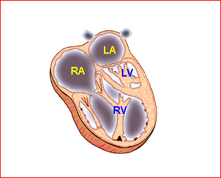
The aorta arises from the right ventricle and lies anterior to and left of the pulmonary artery, which is connected to the left ventricle and lies posteriorly and medially. Complete TGA is uneventful in utero. Survival after birth depends on the amount and size of the mixing of two otherwise independent circulation systems. In the case of an intact ventricular septum, the newborn presents shortly after birth with cyanosis and deteriorates rapidly.

Fig 1

Incidence: 2 per 10,000 live births, accounting for 5% of CHD.

Sonographic diagnosis:

  • The aorta and pulmonary artery are parallel instead of crossing at the level where they arise from the ventricles.
  • The vessel connected to the left ventricle has a posterior course and bifurcates into two pulmonary arteries.
  • The vessel connected to the right ventricle has a long upward course and gives rise to the brachiocephalic vessels.
  • Other cardiac anomalies, especially VSD or pulmonary stenosis.
  • Atrioventricular block is common.
  • Doppler study may show abnormal pulmonary venous blood flow velocity waveforms, and lower pulsatility indices in the middle cerebral artery.
  • Pitfalls:
    • It can often be confused with double-outlet right ventricle or tetralogy of Fallot.
    • TGA can be missed if the main pulmonary artery is not distinguished from the ascending aorta.
    • In cases of mild or isolated forms or corrected TGA, the screening ultrasound may show only an isolated VSD or a small VSD, with a tiny pulmonary trunk.
  • Usually diagnosed after 16-18 weeks, but possible after 13-15 weeks with transvaginal ultrasound in some cases.
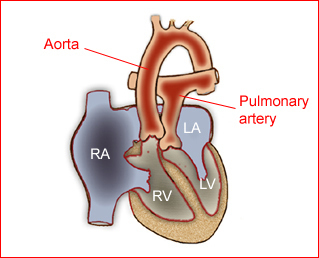
Fig 1:  Schematic drawing of transposition of the great arteries (LA = left atrium, LV = left ventricle, RA = right atrium, RV = right ventricle)

Video clips of Transposition of the Great Arteries (TGA)
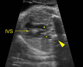
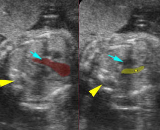
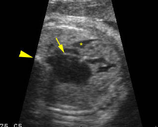
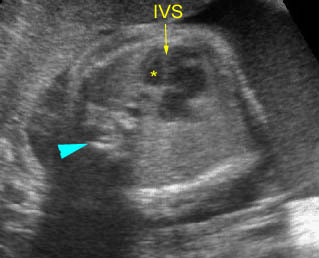
Transposition of the great vessels:  Long-axis view: aortic root (solid circle) and pulmonary trunk (*) parallel arising from right and left ventricle respectively (arrowhead = spine)
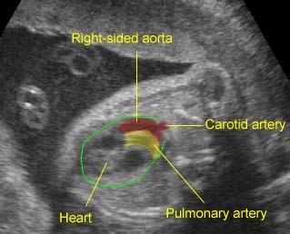
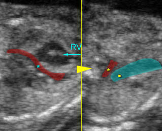
Transposition of the great arteries:  Long axis view: the right-sided aorta running parallel to the left-sided pulmonary artery. Note: the carotid artery arising from the aortic arch

Associations: Other cardiac defects are seen in 50% of cases.

Management: Careful prenatal and postnatal search for associated anomalies and karyotype (including FISH for chromosome 22q11 deletion) are indicated. Delivery should be performed where immediate pediatric cardiac consultation is available.

Prognosis: Complete TGA is well tolerated in utero. Postnatal surgery improves the prognosis, however, the mortality rate is still high overall, especially when associated with other abnormalities. Prenatal diagnosis may improve the preoperative condition of the neonates.

Read More

Ebstein’s Anomaly

Ebstein’s Anomaly

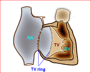
Ebstein’s anomaly is characterized by the congenital downward displacement of the tricuspid valve on the interventricular septum and posterior displacement of the valve with severe dysplasia of the right ventricle. As a result, the right atrium is typically massively enlarged. The valvular orifice is formed within the ventricular cavity at the junction of the atrialized inlet and functional ventricular components.

Fig 1

Incidence: Rare, accounting for only 1% of cardiac anomalies.

Sonographic findings:

Fig2

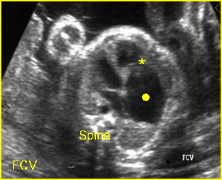
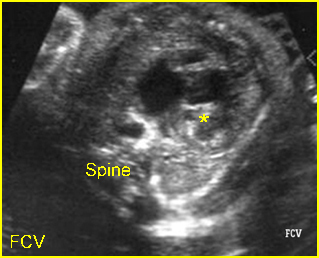
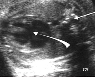
  • Low insertion of the tricuspid valve, demonstrated in FCV.
  • The right atrium is unusually enlarged.
  • Small right ventricle or dysfunction.
  • Doppler study may often show some degree of tricuspid regurgitation, possibly leading to hydrops fetalis.
  • Normal FCV excluding the possibility of Ebstein’s anomaly.
  • The normal reference range of the mitral valve-tricuspid valve distance has been described for each gestational week, allowing identification of abnormal downward displacement of the medial tricuspid cusp in the case of Ebstein’s anomaly.
  • Differential diagnosis:
    • Pulmonary atresia with infarct ventricular septum.
    • Tricuspid valve regurgitation can occur in 6-7% of normally grown fetuses, but the valve morphology and size are normal.
    • Uhl’s anomaly (right anterior ventricular wall dilated due to the defect of cardiac muscle) of the heart mimicking Ebstein’s anomaly.
  • Usually diagnosed after 16-18 weeks

Fig 1:  Schematic drawing of Ebstein anomaly showing downward displacement of the tricuspid valves (RA = right atrium, RV = right ventricle, TV = tricuspid valves)

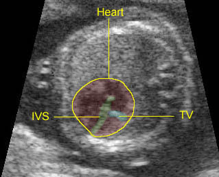
Fig 2:  Ebstein anomaly   Four-chamber view: enlarged atrium (solid circle) with low attachment of tricuspid valve (*)

Video clips of Ebstein’s anomaly

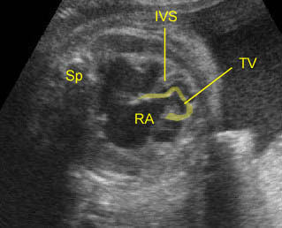
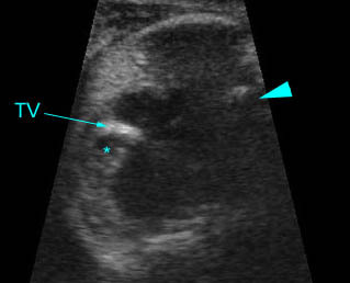
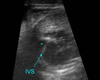
Ebstein’s anomaly:  Extremely low insertion of the tricuspid valve (TV) with marked cardiomegaly (IVS = interventricular septum, RA = right atrium, Sp = spine)

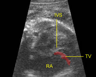
Ebstein’s anomaly:  Low insertion of the tricuspid valve (TV) with marked cardiomegaly (IVS = interventricular septum)

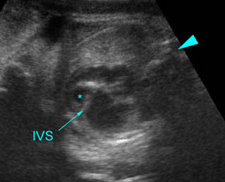
Ebstein’s anomaly:  Ebstein’s anomaly in case of corrected transposition of great arteries: Low insertion of the tricuspid valve (TV) of the left-sided right ventricle (IVS = interventricular septum)

Associations: Other cardiac defects; e.g. pulmonary stenosis, ASD, TOF, TGA.

Management: Termination may be offered in the case of fetuses with associated anomalies or pulmonary atresia or severe tricuspid regurgitation with enlarged right ventricle. In continuing pregnancies, serial ultrasound examinations should be performed and delivery should be performed where immediate pediatric cardiac consultation is available.

Prognosis: Poor in most cases but better in mild cases with appropriate surgical correction.

Read More

Hypoplastic Right Heart Syndrome (HRHS)

Hypoplastic Right Heart Syndrome (HRHS

HRHS is characterized by a small right ventricle, with pulmonary valve atresia with intact ventricular septum.

Fig 1

Incidence: Rare, accounting for 1.7% of structural heart defects.

Sonographic findings:

 Fig 2, Fig 3, Fig 4

  • Small right ventricle on the FCV.
  • Atresia of the pulmonic valve.
  • Intact ventricular septum.
  • The tricuspid valve may be small but patent thus distinguishing HRHS from triscuspid atresia.
  • Doppler imaging usually shows minimal or no flow across the tricuspid valve or sometimes tricuspid valve regurgitation, with reversal of flow in the ductus arteriosus/pulmonary artery.
  • An atrial septal defect is usually present.

 Associations: Other defects of the heart, not related to extracardiac anomaly or chromosome abnormalities.

Management: Careful prenatal and postnatal search for associated anomalies is required. Termination of pregnancy should be considered before viability.

Prognosis: Overall poor, but better than that of HLHS. However, the mortality rate is still high. Surgical correction is appropriate in some selected cases.

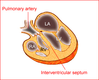
Fig 1:  Schematic drawing of  tricuspid atresia; Note small right atrium (RA) and right ventricle (RV) and VSD (LA = left atrium, LV = left ventricle)

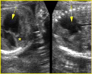
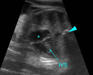
Fig 2:  Hypoplastic right heart syndrome  Very small right ventricle (*)

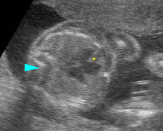
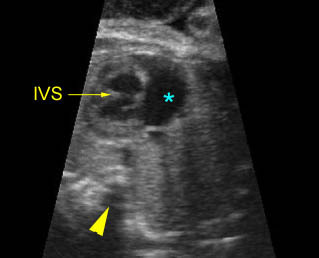
Fig 3:  Hypoplastic right heart syndrome   Left : Four-chamber view: small right ventricle (*) with common atrium

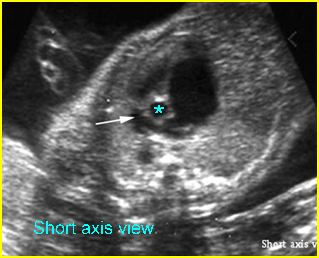
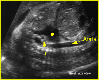
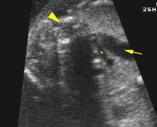
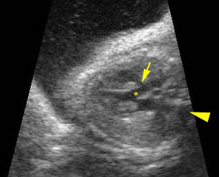
Fig 4:  Hypoplastic right heart syndrome  Short-axis view: small pulmonary trunk (arrow) compared to aorta (*) with large atrium

Video clips of Hypoplastic left heart syndrome 

Hypoplastic right heart:  Long-axis view: small right ventricle and small pulmonary trunk (*) compared with ascending aorta (arrow) (arrowhead = spine)

Triscuspid atresia:  Four-chamber view: extremely small right ventricle (*) with VSD, thickened and echogenic tricuspid valve (arrowhead = spine)

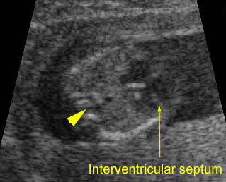
Tricuspid atresia:  Four-chamber view: small right ventricle (*), with thickened and echogenic tricuspid valve (IVS=interventricular septum)

Tricuspid atresia:  Four-chamber view: small right ventricle (arrow) with interventricular septum defect (*),arrowhead = spine

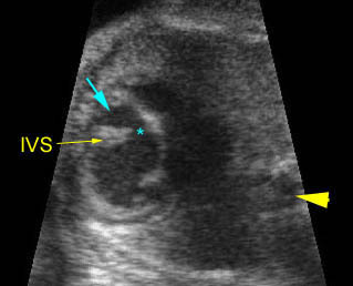
Tricuspid atresia:  Four-chamber view: small right ventricle (*), with thickened and echogenic tricuspid valve (IVS=interventricular septum arrowhead = spine)

Read More

Hypoplastic Left Heart Syndrome (HLHS)

Hypoplastic Left Heart Syndrome (HLHS)

HLHS is characterized by a very small left ventricle, with mitral and/or aortic atresia. Blood flow to the head is supplied by retrograde flow through the ductus arteriosus.

Fig 1

Incidence: 1 in 3000 births, accounting for 10-15% of neonatal deaths from cardiac causes.

Sonographic findings:

Fig 2, Fig 3

  • Small left ventricle on the FCV.
  • Hypoplasia of the mitral and aortic valves.
  • Ascending aorta is often hypoplastic.
  • The movement of the mitral valve is severely impaired.
  • Atrial septal defect.
  • Retrograde flow within the ascending aorta and aortic arch, from ductus arteriosus.
  • Abnormal Doppler pattern of pulmonary venous flow which appears to be a reliable predictor of restriction of the atrial septum in the neonate.
  • Congestive heart failure and hydrops fetalis can occur in cases with tricuspid regurgitation.
  • Increased nuchal translucency thickness at 10-14 weeks of gestation.
  • Pitfalls:
    • Mild hypoplastic left heart often occurs with aortic coarctation of aorta.
    • The left side of the heart can be nearly normal in size in the early second trimester and become severely hypoplastic in late pregnancy.
    • Functional aortic stenosis may show retrograde flow in the aortic arch like HLHS.
  • Usually diagnosed in the second half of pregnancy, though possible as early as 13 weeks (with a persistent reversed flow of ductus venosus during atrial contraction).

Fig 1:  Schematic drawing of hypoplastic left heart syndrome (LA = left atrium, LV = left ventricle, RA = right atrium, RV = right ventricle)

Fig 1:  Hypoplastic left heart syndrome  Very small left ventricle (*)

Fig 3:  Hypoplastic left heart syndrome   Short axis view: markedly small aortic root (arrow) (solid circle = right atrium)

Video clips of Hypoplastic left heart syndrome 

HLHS / Arrhythmias :  Four-chamber view: very small left heart with thickened ventricular wall with irregular heart beats, arrow = flap of foramen ovale, * = pericar-dial effusion, arrowhead = spine

Hypoplastic left heart syndrome :  Four-chamber view: single ventricle (right) (*),arrowhead = spine

Hypoplastic left heart syndrome:  Arch view: enlarged ductal arch (arrow) and small aortic arch (*), arrowhead = spine

Hypoplastic left heart syndrome :
– Long-axis view: small aortic root (*) arising from small left ventricle (arrowhead = spine)
– Arch view: small aortic arch (*), compared to large ductal arch (arrowhead = spine)

Hypoplastic left heart syndrome :  Four-chamber view: small left ventricle (*), compared to right ventricle (arrowhead = spine)

Associations: Coarctation of aorta, with chromosome abnormalities seen in 12-25% of cases, especially microdeletion 22q11, trisomy 18 or Turner syndrome.

Management: Careful prenatal and postnatal search for associated anomalies is required as well as chromosome study. Termination of pregnancy should be offered before viability. In continuing pregnancy, delivery should occur where immediate pediatric cardiology and surgery support are available. Vaginal delivery can be allowed. The proportion of infants with HLHS treated with staged surgical palliation and infant heart transplant has increased, resulting in major improvements in survival and quality-of-life outcomes.

Prognosis: Very poor, but better with staged reconstruction surgery in selected cases (survival rate of 25-40%).

Recurrence risk: About 2%, but higher in rare autosomal recessive cases.

Read More

Atrioventricular Canal Defect

Atrioventricular Canal Defect

AVC defects include a spectrum of lesions ranging from an isolated primum to complete defects of the atrioventricular canal, commonly referred to as endocardial cushion defect.

The defect causes a spectrum of atrioventricular outlet arrangements. In the complete form, persistent common atrioventricular canal, the tricuspid and mitral valve are fused in a large single atrioventricular valve that opens above the bridges of the two ventricles. This valve has an anterior and a posterior leaflet. In the incomplete form, various amounts of tethering of one or both leaflets to the crest of the interventricular septum lead to a connection that creates functional atrial or ventricular septal defects, ventriculoatrial septal defects or valvular abnormalities.

Incidence: AVC accounts for 7% of structural heart defects.

Sonographic findings:

 Fig 1

  • Complete defect is usually easy to diagnose by demonstrating an obvious deficiency of the central core structures of the heart on FCV.
  • Increased nuchal translucency thickness at 10-14 weeks of gestation and nuchal translucency can be a useful early screening for CHD.
  • The atria may be dilated as a consequence of atrioventricular insufficiency.
  • The incomplete forms are more difficult to recognize. The tricuspid and mitral valve can attach at the same level at the crest of the septum. This apical displacement of the mitral valve elongates the left ventricular outflow tract.
  • Color Doppler facilitates the visualization of the central opening of the single atrioventricular valve; the outcome is adversely affected when the AV valve is regurgitant.
  • The atrial septal defect is of the ostium primum type (because the septum secundum is not affected) and thus is close to the crest of the interventricular septum.
  • Though it is detectable by FCV, the detection rate is currently <50%. Live 3D ultrasound may be helpful.
  • Usually diagnosable after 16-18 weeks but possible after 10-14 weeks with transvaginal ultrasound.
  • Pitfalls:
    • False dropout can occur from apical FCV, therefore, it is important to image the defects in a scan plane perpendicular to the septum.
    • A dilated coronary sinus may be mistaken for primum ASD.

Fig 1:  Atrioventricular canal  Four-chamber view: common atrium and ventricle, no visible central crux (arrow)

Video clips of Atrial Septal Defect (ASD)

Endocaridal cushion defect:  Four-chamber view: atrial and ventricular septal defect, arrowhead = spine

Endocardial cushion defect :  Four-chamber view: abnormal cardiac axis, atrial septal and ventricular septal defect (arrowhead = spine)

Associations: Chromosome abnormalities in more than 50% of cases with 60% being trisomy 21 and 25% being trisomy 18, also including extracardiac defects and heterotaxy syndrome in particular. Isolated AVC is a higher risk for trisomy 21 than that with associated anomalies.

Management: Careful prenatal and postnatal search for associated anomalies is required. All continuing pregnancies should be karyotyped. Serial sonography should be performed.

Prognosis: Poor when associated with chromosome abnormalities or other anomalies. Isolated AVC can be repaired with a survival rate of more than 80%.

Read More

Ventricular Septal Defect (VSD)

Ventricular Septal Defect (VSD)

VSD is an opening of the ventricular septum causing communication between the two ventricles. Most (80% of cases) are perimembranous defects.

Incidence: VSD is the most common cardiac defect, accounting for nearly one-third of all heart defects.

Sonographic findings:

Fig 1, Fig 2

  • Perimembranous and subaortic VSD are best visualized with a left outflow tract view and best imaged when the ultrasound beam is perpendicular to the focused septum.
  • Muscular defects, which are difficult to observe, are best searched for in the short-axis view by trying to demonstrate a connection between the two ventricles.
  • Color Doppler is helpful in demonstrating shunting across the septum.
  • Color Doppler spatiotemporal image correlation has the potential to simplify visualization of the location and extent of ventricular septal defects.
  • Pitfalls:
    • Defects smaller than 1-2 mm will escape detection.
    • False dropout is most likely to occur when the ultrasound beam is parallel to the defects.
    • Isolated VSD is one of the common defects rarely diagnosed correctly, especially on routine screening at 18 weeks.
  • Usually diagnosable between 16 and 18 weeks, but diagnosis possible at 14 weeks with transvaginal ultrasound.
  • Some cases may show thickened NT between 11 and 14 weeks.

Large ventricular septal defect:  Four-chamber view: incomplete interventricular septum (*), (arrowhead = spine)

ASD & VSD:  Atrial septal and ventricular septal defect
Four-chamber view: abnormal cardiac axis, no atrial septum (*) and incomplete ventricular septum (arrowhead = spine)

Ventricular septal defect:  Long axis view: small interventricular defect (arrow) (not seen in the four-chamber view) (* = aortic root, arrowhead = spine)

Associations: May occur alone or be associated with certain syndromes or other heart abnormalities including tetralogy of Fallot and transposition of the great vessels. VSD is also associated with increased risk of aneuploidy, even seemingly isolated VSD.

Management: Careful prenatal and postnatal search for associated anomalies is required. All continuing pregnancies should be karyotyped. Isolated VSD should not alter standard obstetric care.

Prognosis: Usually good for isolated and mild forms, but poor when associated with associated defects and large defects, especially the prenatally diagnosed defects. Ventricular septal defect can undergo spontaneous closure in utero, especially defects of <3 mm.

Recurrence risk: Most are multifactorial with a recurrent risk of 2-4%.

Read More

Atrial Septal Defect (ASD)

Atrial Septal Defect (ASD)

ASD is an opening of the atrial septum causing communication between the two atria. Embryologically, the first septum (primum) to develop arises from the endocardial cushion and starts at the AV valves, growing toward the free wall of the combined atrium. Later a second septum (secundum) starts at the free wall of the atrium and grows toward the septum primum. The gap between the two is called the ostium primum. When the fusion between the septum primum and endocardial cushion has occurred, the septum primum fenestrates. The resulting communication is called ostium secundum. A second septum then extends on the right side of the septum primum and covers part of the ostium secundum. The remaining orifice is the foramen ovale, and the foramen ovale flap is the lower part of the septum primum. Thus, defects that are close to the endocardial cushion are called the ostium primum, and defects in the area of the foramen ovale are the ostium secundum. The primum ASD is the simplest form of atrioventricular septal defect or partial AV canal.
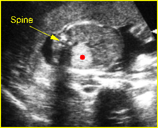
Fig 1

Incidence: ASD accounts for 8% of structural heart defects; secundum defects are the most common and are found in 0.07 per 1000 infants.

Sonographic findings:

    Fig 2, Fig 3

  • Secundum ASDs are most commonly isolated.
  • Occasionally, ASD is related to other cardiac lesions including mitral, pulmonary, tricuspid or aortic atresia.
  • Abnormal oscillatory pattern of the foramen ovale flap motion by M-mode.
  • Pitfalls:
    • Prenatal diagnosis of secundum ASD is relatively difficult due to the physiologic presence of the foramen ovale. Most likely, only unusually large defects can be recognized with certainty.
    • The detection rate was poor for isolated septal defects but higher when associated with other complex lesions or abnormal four-chamber views

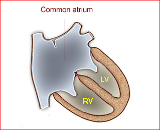
Fig 1:  Schematic drawing: Atrial septal defect with common atrium (RV = right ventricle, LV = left ventricle)

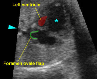
Fig 2:  Atrial septal defect (ASD)   Four-chamber view: common atrium (*) no atrial septum and flap of foramen ovale

Fig 3:  Atrial septal defect (ASD)  Four-chamber view: common atrium, no atrial septum and flap of foramen ovale

Video clips of Atrial Septal Defect (ASD)

Atrial septal defect:  Four-chamber view: abnormal cardiac axis, no atrial septum seen (arrowhead = spine)

ASD & VSD:  Atrial septal and ventricular septal defect
Four-chamber view: abnormal cardiac axis, no atrial septum (*) and incomplete ventricular septum (arrowhead = spine)

ASD occurs in the setting of other defects:

  • Ebstein’s anomaly
  • Hypoplastic right heart
  • Hypoplastic left heart
  • Pulmonic stenosis
  • Truncus arteriosus

 Associations: Most cases of secundum ASD are isolated but may sometimes be found to be part of syndromes such as Holt-Oram syndrome. The primum defects are more associated with other anomalies. (See the section “Atrioventricular canal defect”.)
Management: Careful prenatal and postnatal search for associated anomalies is required; small defects may become smaller or close after birth, however, moderate to large defects often need surgical correction or catheterization closure devices.
Prognosis: ASDs are not a cause of impairment of cardiac function in utero because a large right-to-left shunt at the level of the atria is a physiologic condition in the fetus. Most affected infants are asymptomatic even in the neonatal period.
Read More

Pulmonary Sequestration

Pulmonary Sequestration

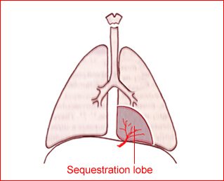
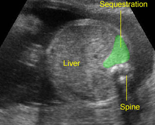
Pulmonary sequestration (PS) is a supernumerary lobe of the benign pulmonary tissue, separated from the normal tracheobronchial tree, having its own vascular supply, which usually arises from the thoracic or abdominal aorta. The lesions are pathologically divided into intralobar and extralobar forms (invested by its own pleura). Intralobar sequestrations almost always drain into pulmonary veins whereas most extralobar lesions drain into systemic veins. Approximately 10-15% of extralobar forms are found within or below the diaphragm, usually on the left.

Incidence: 1 in 1000 births.

Fig 1

Sonographic findings:

   Fig 2,  Fig 3,  Fig 4

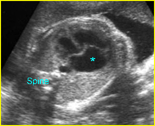
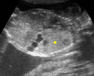
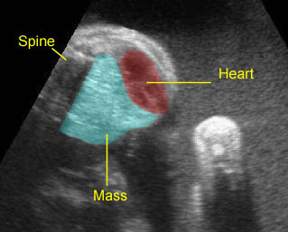
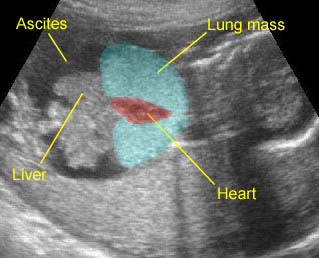
  • Typically homogeneous echogenic mass varying in size in the chest (90% of cases) or abdomen (10% of cases).
  • Unilateral in most cases, usually left lower lobe.
  • Mediastinal shift, pleural effusion and hydrops fetalis if the mass is large enough.
  • Visualization of a feeder vessel arising from the thoracic or abdominal aorta feeding the mass strongly suggestive of extralobar sequestration.
  • Regression on the follow-up scans (decreasing in size, echogenic, and mediastinal shift) is often seen.
  • Main differential diagnoses are other lung masses, including type III CCAM, CDH, bronchial atresia, and mediastinal teratoma. The extrathoracic form may be differentiated from neuroblastoma, renal tumor, and teratoma. PS usually is echogenic, left-sided, and can be identified in the second trimester whereas neuroblastoma is most often cystic, right-sided, and identified in the third trimester.
  • Pitfalls:
    • A feeding vessel of pulmonary sequestration in the thorax is not always seen, therefore it is often indistinguishable from Type III CCAM.
    • Thoracoabdominal masses including CCAM, CDH, neuroblastoma, lymphangioma, and bronchial atresia, mimicking PS.
    • The extrathoracic type is easily overlooked.
    • MRI may be helpful in difficult cases.
  • Usually detected in the third trimester but some cases are detected early in the second trimester.

Fig 1:  Schematic drawing: Bronchopulmonary extra-lobar seques-tration in the inferior portion of the thorax

Fig 2:  Extralobar pulmonary sequestration   Cross-sectional scan of thorax: bright echogenic mass (solid circle) anterior to the spine

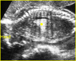
Fig 3:  Extralobar pulmonary sequestration  Sagittal scan of thorax: intrathoracic bright echogenic mass (solid circle) in the shape of lung lobe

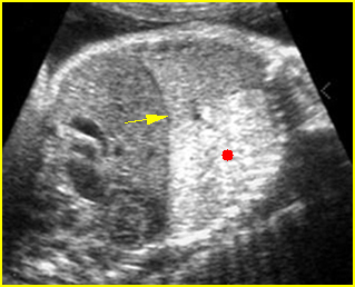
Fig 4:  Extralobar pulmonary sequestration   Oblique sagittal scan of the fetal trunk: markedly echogenic, lung-shaped mass in the chest (solid circle) (arrow = intact diaphragm)

Video clips of pulmonary sequestration

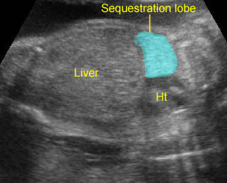
Lung sequestration: Extralobar sequestration: Echogenic lung mass with lobar in shape

Lung sequestration:  Extralobar sequestration in the upper abdomen: Echogenic mass with lobar in shape behind the liver

Associations: Associated abnormalities including cardiac defect, CDH or lung abnormalities are found in 10% of intralobar type cases and 58% of extralobar type cases. Sequestration and CCAM may be seen together, which is suggestive of a common embryogenesis despite diverse morphology.

Management: Conservative and follow-up ultrasound examination is appropriate in most cases. Many extralobar pulmonary sequestrations dramatically decrease in size before birth or after birth. Fetal intervention with a thoracoamniotic shunt may be needed in some hydropic cases. Delivery should be at term in a tertiary center. Cases with residual mass at birth should have surgical resection. An ex utero intrapartum therapy (EXIT) strategy with resection of the mass during the EXIT procedure may be appropriate in selected cases. However, the need for surgery should be based on appropriate postnatal investigations (e.g. CT scans), rather than on antenatal behavior.

Prognosis: Good prognosis in the majority of cases, spontaneous regression occurs in many cases, and the persistent cases are resected safely postnatally; poor if associated with hydrops, though reversible in some cases.

Read More

Congenital Cystic Adenomatoid Malformation (CCAM)

Congenital Cystic Adenomatoid Malformation (CCAM)

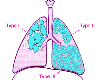
CCAM is a hamartoma of the lung that may involve a whole lung or a single lobe. Multiple cysts replace pulmonary parenchyma. It can also occur in lung sequestration. The cysts are usually unilateral and are classified into three types as follows:

     Fig 1

  • macrocystic (type I): few in number and large, up to 7 cm, and thick-walled
  • mixed (type II): multiple and smaller (about 1 cm)
  • microcystic (type III): very small, <0.5 cm, and not visible as distinct cysts by ultrasound; the lesions appear as bulky, intrathoracic, solid masses with increased echogenicity.

Incidence: Unknown.

Sonographic findings:

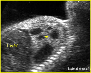
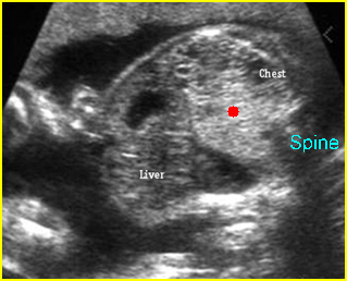
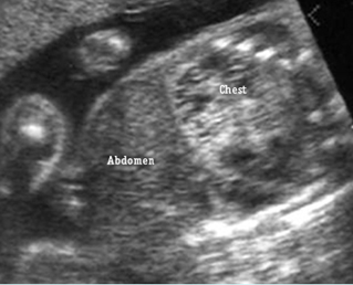
Fig 2, Fig 3, Fig 4

  • Echogenic or sonolucent mass in one side of the chest possibly shifting the heart.
  • Unilateral in most cases.
  • Hydrops fetalis (<10% of cases), commonly seen with a large mass, in particular a greater CAM volume ratio.
  • Polyhydramnios (65% of cases) secondary to esophageal obstruction from the intrathoracic mass, obstructed venous return or decreased absorption of lung fluid.
  • Doppler study reveals a normal blood supply, unlike the sequestration.
  • Main differential diagnosis:
    • If cystic mass: CCAM, CDH, bronchogenic or neuroenteric cyst, esophageal duplication
    • If solid mass: microcystic CCAM, pulmonary sequestration, CHD.
  •  Pitfalls:
    • Most often confused with congenital diaphragmatic hernia.
    • Acoustic enhancement posterior to the heart mimicking microcystic mass.
    • The normal thymus may occasionally simulate hypoechogenic mass.
  • First diagnosable between 12 and 18 weeks of gestation

Fig 1:  Schematic drawing: The three type of CCAM: type I (large cysts of variable size), type II (small cystic type), and type III (solid appearance)

Fig 2:  Cystic adenomatoid malformation   Oblique sagittal scan of the fetal trunk: solid-cystic mass (*), mostly cystic, in the left chest (CCAM type I)

Fig 3:  Cystic adenomatoid malformation   Oblique sagittal scan of the fetal trunk: echogenic mass in the left chest (solid circle)

Fig 4:  Cystic adenomatoid malformation  Oblique sagittal scan of the fetal trunk: partial solid and partial cystic mass in the left chest (CCAM type II)

Video clips of congenital cystic Adenomatoid malformation (CCAM)

CCAM (Congenital cystic adenomatoid malformation; CCAM)  Coronal scan of the chest: large echogenic mass (solid circle) in the left thorax with some cystic changes

CCAM (Congenital cystic adenomatoid malformation: CCAM)   CCAM type III: Large echogenic lung mass with mediastinal shift

Laryngeal Atresia :  Bilateral enlarged echogenic lung caused by high airway obstruction, associated with ascites, simulating bilateral CCAM type III

Associations: Usually not associated with other anomalies.

Prognosis: Good prognosis in the majority of cases especially the cystic type and it can spontaneously regress antenatally, but can not continue after birth; poor prognosis if associated with hydrops, especially microcystic type, lung hypoplasia, prematurity or severe associated malformations.

Management: Follow-up ultrasound examination is required, with no active intervention in the absence of acute polyhydramnios or hydrops. Persistent CCAM needs postnatal surgical removal.

Recurrence risk: Unknown.

Read More

Diaphragmatic Hernia

Diaphragmatic Hernia

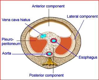
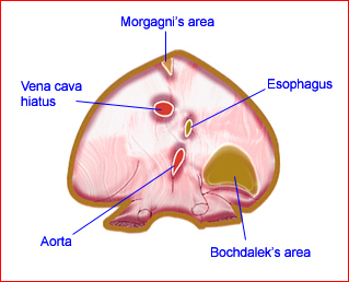
Diaphragmatic hernia (CDH) is a herniation of abdominal viscera into the fetal chest, which results from a congenital defect in the fetal diaphragm. The most common location is posterolateral (Bockdalek) on the left side (85-90%) followed by a retrosternal (foremen of Morgagni hernia) defect.

Incidence: 1 in 2500-4000 chromosomally normal fetuses.

Fig 1, Fig 2, Fig 3

Fig 1:  Schematic drawing: Developmental components of the diaphragm

Fig 2:  Schematic drawing of the diaphragm seen from below; note the location of potential site of Bochdalek’s hernia

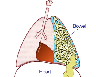
Fig 3:  Schematic drawing: Diaphragmatic hernia: bowel loops in the chest with mediastinal shift

Sonographic findings:

Fig 3, Fig 4, Fig 5, Fig 6

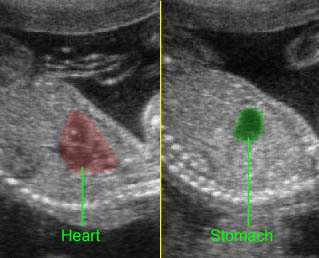
  • Abdominal organs above diaphragm, visualization of peristalsis.
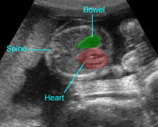
  • Mediastinal shift, heart displacement to the right side.
  • 90% of left CDH detected due to visualization of displaced heart and stomach in the chest.
  • Unilateral in most cases, bilateral in <5% of cases.
  • Undetected fluid-filled stomach below the diaphragm.
  • Generally, abdominal circumference not significantly different from normative data.
  • Thickened nuchal translucency at 10-14 weeks in nearly 40% of cases.
  • MRI and CT amniography are helpful if obesity or fetal position make detection difficult.
  • Three-dimensional ultrasound is helpful in the assessment of lung volume in cases of CDH, and is a potential predictor for pulmonary hypoplasia and postnatal outcome. The lung to head ratio may be a good predictor for fetal outcome.
  • Main differential diagnoses include:
    • Cystic adenomatoid malformation (intact diaphragm)
    • Congenital hiatal hernia (midline dilated tubular structure)
    • Pulmonary sequestration (intact diaphragm)
    • Dextrocardia (unlike CHD, the apex points to the right)
    • Lung tumor (dilated tubular structures).
  • Right-sided hernia (<10% of cases):
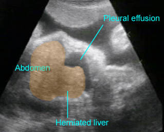
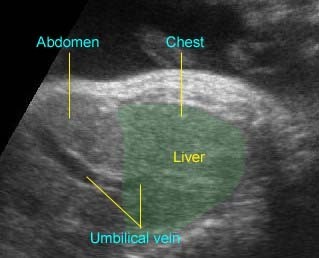
  • Right-sided chest mass, portal vein and gallbladder can be tracked within the mass
  • The liver alignment is changed
  • Pleural effusion is often seen on the same side
  • The fluid-filled stomach is usually in a relatively normal location
  • MRI is helpful in demonstration of the liver in the chest.
  • Pitfalls:
    • Despite high accuracy of sonographic diagnosis, a significant number of cases are missed. More than half are related to failure in following established guidelines.
    • The classic sonographic features are not always present at the time of initial evaluation, therefore, CDH should be differentiated in all echogenic chest masses.
    • Differentiating between congenital diaphragmatic eventration (better prognosis) and CDH may be very difficult.
    • A collapsed bowel in the chest can easily be overlooked.
    • Right-sided hernia is often more subtle.
    • It may be confused with congenital hernia, a midline dilated tubular structure in the thoracic cavity.
    • Lung changes are easily overlooked, especially if the stomach is in the abdomen.
  • Diagnosable as early as 12 weeks for left-sided hernia and 17-18 weeks for right-sided hernia

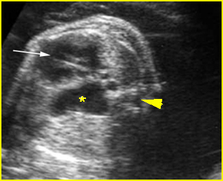
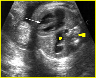
Fig 4:   Pseudo-diaphragmatic hernia  Oblique cross-sectional scan of the thorax: the stomach (*) seems to be located in the chest due to tangential scan (arrow = heart)

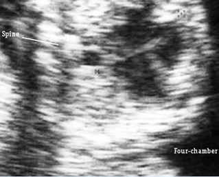
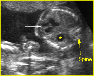
Fig 5:  Diaphragmatic hernia  Cross-sectional scan of thorax at the level of four-chamber view: cystic structure of bowel loop (*) located in the chest with cardiac displacement (arrow) to the right (arrowhead = spine)

Fig 6:  Diaphragmatic hernia   Cross-sectional scan of thorax at the level of four-chamber view: cystic structure of bowel loop (solid circle) located in the chest with cardiac displacement to the right (arrowhead = spine)

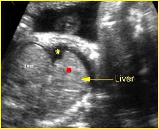
Fig 7:  Right diaphragmatic hernia   Sagittal scan of the chest: part of liver (solid circle) upward displacement to the chest (* = pleural effusion)

Video clips of diaphragmatic hernia

Diaphragmatic hernia:  Sagittal scan: The stomach is herniated into the chest, next to the heart

Diaphragmatic hernia:  Cross-sectional scan: The stomach is herniated into the chest, not seen in the abdomen, with cardiac displacement

Diaphragmatic hernia:  Cross-sectional scan: The stomach is herniated into the chest with cardiac displacement

Right-sided diaphragmatic hernia:  A portion of the liver herniated into the chest with pleural effusion

Right-sided diaphragmatic hernia:  Right-sided diaphragmatic hernia: the liver herniated in the chest, the umbilical vein running into the chest

Associations: More than 25% had associated anomalies, including anomalies of the heart (20%), brain (30%), genitourinary system, craniofacial region, or limbs. Chromosomal abnormalities occur in 5-15%, with the most common being trisomy 18.

Management: In utero tracheal occlusion to promote lung growth in severe cases was impressive in preliminary studies but was not confirmed by later randomized control trial (RCT). Intrauterine repair should be at present considered as an investigational technique. Ex utero intrapartum treatment has been successful in a few cases. The delivery should be in a tertiary center. CDH needs effective immediate resuscitation and maintaining adequate gas exchange by mechanical ventilation and surgery is delayed until the respiratory status is stable. Postoperative extracorporeal membrane oxygenation (ECMO) is helpful in decreasing the mortality rate.

Prognosis: Poor; the main two poor prognostic factors are pulmonary hypoplasia and associated anomalies; better prognosis is reported for isolated cases with proper surgical correction, especially with ECMO treatment, as well as high-frequency ventilation, and antenatal steroids for lung maturity.

Recurrence risk: Usually sporadic with a low recurrence risk but a familial case with a higher risk for recurrence was found in <2% of cases.

Read More