Amniotic Band Syndrome (ABS)

Amniotic Band Syndrome (ABS)

ABS is a destructive fetal complex caused by disruption of the amnion. It may be a sequela of intrauterine rupture of the amnion, resulting in oligohydramnios and passage of the fetus into the chorionic cavity, causing fetal compression and localized fetal ischemia, possibly resulting in a pathogenic mechanism of extremely variable malformations. A focal development error of connective tissue may be involved. The malformations range from minor constriction rings and lymphedema of the digits to complex, bizarre multiple anomalies that are attributed to amniotic bands that stick, entangle and disrupt fetal parts, particularly the cranium base, spine, or extremities.

Incidence: 1 in 1200 to 1 in 15,000 live births.

Sonographic findings:

  • A wide variety of anomalies, including neural tube defect, unusual asymmetric or bizarre clefts of the lip/palate, abdominal wall defects, and amputation-type defects.
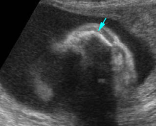
  • Constriction rings of the limbs, typically edema distal to the constriction ring.
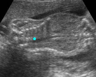
  • Amputation defect of the extremity.
  • Craniofacial defects such as acrania, craniosynostosis.
  • Unusual pattern of facial clefting.
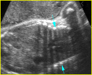
  • Occasionally, amniotic bands themselves are visualized.
  • The differential diagnosis depends on the body part involved. For example, cranial defects caused by ABS must be differentiated from acrania or cephaloceles. Abdominal wall defects secondary to ABS should be differentiated from limb-body wall complex (LBWC). Focal musculoskeletal anomalies should be differentiated from other causes such as mesomelic dysplasia, radial ray defects, etc.. Multisystem abnormalities favor the diagnosis of ABS.
  • Usually first diagnosable in the second trimester.
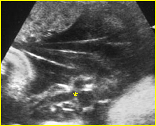
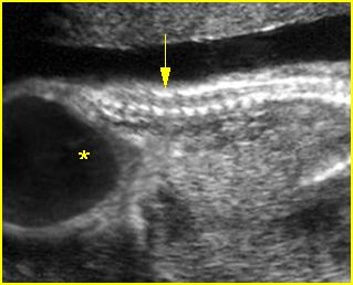
Fig 1:  Amniotic bands  Several amniotic bands (arrows) in amniotic cavity (* = umbilical cord)
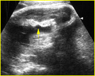
Fig 2:  Amniotic band syndrome   Constricted ring on the forearm of the fetus with amniotic band syndrome (arrow)
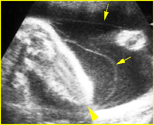
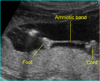
Fig 3:  Amniotic band syndrome   The lower leg abruptly ends (arrowhead) and surrounded by thin amniotic bands (arrows)

Video clips of amniotic band syndrome (ABS)
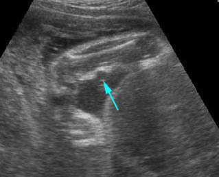
Amniotic band syndrome :  The fetal foot with restricted movement secondary to amniotic band
Amniotic band syndrome :  Constriction ring on the forearm (arrow) secondary to amniotic band
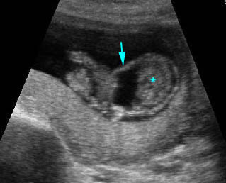
Amniotic band syndrome :  Abnormal band (arrow) surrounding visceral organs (*) outside the body
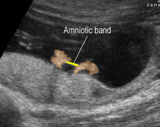
Amniotic band syndrome :  Amniotic band adhered between the both hands

Associations: Non-specific multiple defects.

Management: Depends on the extent of the anomalies. Termination of pregnancy can be offered for severe forms. Expectant management is preferred in most cases of mild forms. Endoscopic release when a limb is at risk of amputation.

Prognosis: Depends on the severity and distribution of anomalies; usually a fatal prognosis for severe craniofacial deformities but a good prognosis for mildly affected infants with isolated limb defects.

Recurrence: Not known to be increased

Read More

Sacral Agenesis

Sacral Agenesis

The spectrum of sacral agenesis and dysgenesis is associated with caudal regression syndrome (CRS) and sirenomelia (mermaid syndrome). Considerable overlap between these abnormalities may exist, with some considering them spectrums of the same entity. A more recent theory, however, suggests that CRS and sirenomelia are pathologically distinct entities.

Caudal Regression Syndrome (CRS)

CRS refers to abnormalities associated with the absence of the sacrum and variable portions of the lumbar spine. This malformative syndrome is mainly seen in cases of maternal diabetes with poor metabolic control.

Incidence: Rare, with an increased frequency in infants of diabetic mothers (200 times, but <1%), or with retinoic exposure in utero.
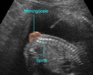
Sonographic findings:

  • Absence of sacrum and variable portions of the lumbar or even thoracic spine.
  • Abrupt interruption of the spine at the dorsal or lumbar level.
  • Abnormal position of the lower limbs.
  • Abnormal pelvic bone, e.g. small, apposed, or absent.
  • The femur bones are of fixed flexion and abduction in a Buddha position.
  • A two-vessel umbilical cord may be seen but this is not as common as with sirenomelia.
  • Increased frequency of renal abnormalities, e.g. agenesis, cystic dysplasia.
  • Oligohydramnios in cases of renal agenesis.
  • Polyhydramnios is occasionally seen if the patient is diabetic.
  • Increased nuchal translucency in late first trimester.
  • Diagnosable in late first trimester, especially with transvaginal ultrasound.
  • The sequence of development of the sacral region ossification is related to gestational age. When the S1-S2 ossification nuclei are visualized at 16-17 weeks of gestation, isolated sacral agenesis can be accurately excluded.

Associations: Non-specific anomalies involving any system may be found.

Management: Termination of pregnancy should be offered. In continuing pregnancy, a multidisciplinary approach is required.

Prognosis: Poor, but in some selected cases a better outcome may be achieved with multidisciplinary management.

Recurrence risk: Rare, however with maternal diabetes or familial history the recurrence risk is higher.

Sirenomelia

Sirenomelia or mermaid syndrome refers to a complex malformation caused by disruptive vascular defects, characterized by a single lower extremity, associated with renal agenesis, and absence of the sacrum. Sirenomelia may be due to an alteration in early vascular development leading to vitelline arterial steal, in which the blood flow is diverted from the caudal region of the embryo to the placenta, resulting in multiple defects of the lower extremities. A single lower extremity in sirenomelia arises from failure of the lower limb bud field to be cleaved into two lateral masses.

Incidence: 1 in 24,000-67,000 births.

Sonographic findings:

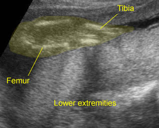
  • Fusion of the lower extremities, the flipper-like deformity of the foot.
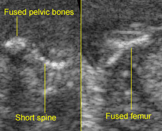
  • Lower spine deformities.
  • Bilateral renal agenesis, leading to oligohydramnios.
  • The fibulas, if present, are between the tibia.
  • The sole of the foot is oriented ventrally instead of dorsally.
  • Single umbilical artery.
  • Situs inversus is frequently seen.
  • Three-dimensional ultrasound is helpful in the diagnosis.
  • Diagnosable in late first trimester, especially with transvaginal ultrasound.
  •  The main differential diagnosis is caudal regression syndrome.

Sirenomelia :  Fusion of the both lower extremities; the both extrremities persistently close to each other

Sirenomelia :  Short spine with fused pelvic bones and Y-shaped fused femur

Associations: Non-specific anomalies involving any system may be found.

Management: Termination of pregnancy should be offered.

Prognosis: Lethal, though an infant with sirenomelia surviving beyond the neonatal period was reported (now 4 years old).

Recurrence risk: Rare, however with maternal diabetes or familial history the recurrence risk is higher.

Read More

Sacrococcygeal Teratoma

Sacrococcygeal Teratoma

A teratoma is generally defined as a tumor arising from pleuripotent cells, composed of a wide variety of tissues haphazardly arranged, usually representing derivatives of their germ layers. Sacrococcygeal teratoma (SCT) is usually obvious at birth, presenting as a large exophytic mass with solid or cystic or mixed components in the sacrococcygeal region. SCT may be classified into four types as follows:

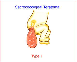
  • Type I: nearly entirely external Fig 1
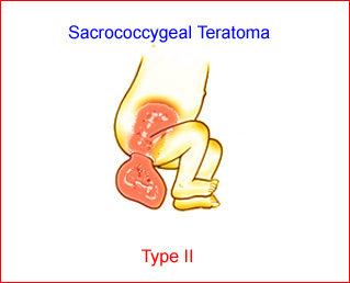
  • Type II: internal and external in equal proportions Fig 2
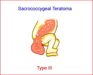
  • Type III: mainly internal Fig 3
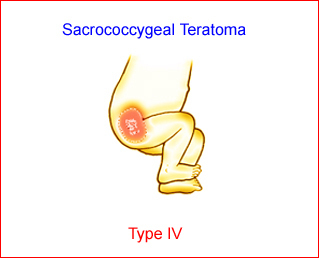
  • Type IV: entirely internal with no external component. Fig 4

Fig 1:  Schematic drawing of SCT type I: predominantly external component

Fig 2:  Schematic drawing of SCT type II: predominantly external component with significant intrapelvic component

Fig 3:  Schematic drawing of SCT type III: predominantly internal component with intra-abdominal extension

Fig 4:  Schematic drawing of SCT type IV: entirely internal component

Moreover, they are histologically categorized into three main groups, including benign (75%), malignant (13%) and immature (12%).

Incidence: SCT is a rare tumor, but the most common fetal tumor, with an incidence of 1:20,000-1:40,000 live births. The male to female ratio is 1:3-4. Most cases are sporadic, but familial presacral teratomas have been reported.

Sonographic findings:

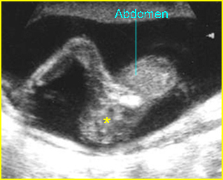
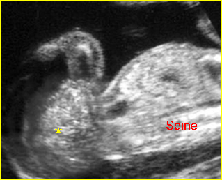
Fig 5, Fig 6, Fig 7, Fig 8, Fig 9, Fig 10, Fig 11

  • The teratomas exhibited three sonographic patterns: mixtures of cystic and solid components in equal proportions, predominantly solid with a few scattered anechoic areas, and unilocular cystic masses. The majority of SCTs are solid or mixed cystic and solid, and only 15% are entirely cystic.
  • Anterior displacement of the bladder in cases of intra-abdominal components.
  • The main differential diagnosis includes
    • anterior or posterior meningomyeloceles
    • conjoined twins, especially with large tumors
    • hemangioma
    • neuroectodermal cyst.
  • The entirely intra-abdominal cystic mass must be distinguished from ovarian cyst, meconium pseudocyst or enteric duplication cyst.
  • Evidence of high-output heart failure, placentomegaly, polyhydramnios, or hydrops fetalis is often seen in large tumors. Doppler echocardiography to detect the presence of congestive heart failure may allow well-timed therapeutic interventions.
  • Umbilical artery waveform or reversed diastolic flow suggest impending fetal demise.
  • Serial ultrasound examinations should be performed to assess amniotic fluid volume, tumor growth, fetal well-being, and early evidence of hydrops.
  • Visualization of an intra-abdominal component, including pelvic structures and vasculature demonstrated with 3D ultrasound, is of prognostic importance. However, there was no correlation between the sonographic appearance and the presence of immature or malignant components.
  • Usually diagnosed in the second and third trimesters.

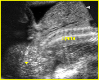
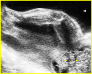
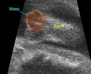
Fig 5:  Sacrococcygeal teratoma   Sagittal scan of the spine (21 weeks): abnormal complex solid mass with heterogeneous echodensity (*) located at the end of sacrum

Fig 6:  Sacrococcygeal teratoma   Sagittal scan of the spine (29 weeks): abnormal complex solid mass with heterogeneous echodensity (*) located at the end of sacrum

Fig 7:  Sacrococcygeal teratoma   Sagittal scan of the spine (27 weeks): abnormal complex solid mass with heterogeneous echodensity (*) located at the end of sacrum

Fig 8:  Sacrococcygeal teratoma   Sagittal scan of the spine (26 weeks): abnormal complex solid mass with heterogeneous echodensity (*) located at the end of sacrum

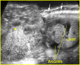
Fig 9:  Sacrococcygeal teratoma  Sagittal scan of the spine (35 weeks): bizarre complex solid mass with heterogeneous echodensity (*) located at the end of sacrum (arrow) and complicated with ascites

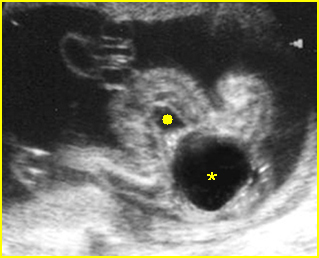
Fig 10:  Sacrococcygeal teratoma   Cross-sectional scan of the lower spine (17 weeks): cystic mass (*) located between bladder (solid circle) and sacrum

Fig 11:  Sacrococcygeal teratoma   Sagittal scan of the lower spine (29 weeks): cystic mass (*) located at the end of sacrum (arrow)

Video clips of sacrococcygeal teratoma

Sacrococcygeal Teratoma :  Mass on the back: the complex cyst at the caudal end of the spine

Associations: Musculoskeletal abnormalities (most common), and renal, cardiovascular, gastrointestinal and CNS anomalies (less common).

Management: The fetuses with a large tumor should be delivered by cesarean section to avoid dystocia and catastrophic hemorrhage during delivery. Small teratomas as well as most cystic tumors allow vaginal delivery after having been punctured previously. Fetal surgical intervention, such as resection, radiofrequency ablation, thermocoagulation, intrauterine shunting in the case of bladder obstruction or percutaneous drainage or aspiration of the cystic type, has proven successful in highly selected cases.

Prognosis: Hydrops fetalis, premature delivery, type III and IV, diagnosis at an earlier gestational age predicts a poor outcome. Types I and II with appropriate surgical correction are related to a better prognosis.

Read More

Spina Bifida

Spina Bifida

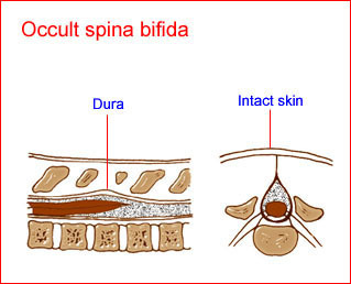
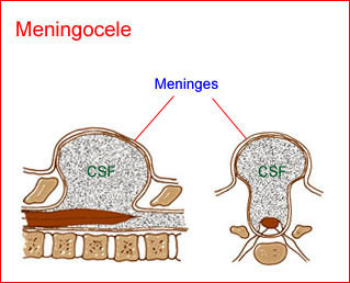
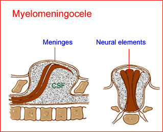
Spina bifida is a defect resulting from failure of closure of the posterior neuropore, occurring anywhere along the spinal axis, but most commonly involving the lumbosacral region (63% of cases). Exposure of neural elements to amniotic fluid and meconium may lead to severe functional impairment. Spina bifida can be categorized as (1) meningocele (sac with CSF only), usually occurring at the upper and lower ends of the neural axis (i.e. occipital, cervical, and low sacral positions), or (2) myelomeningocele (protrusion of a sac containing CSF and neural elements), typically located at the lower thoracolumbar, lumbar, and lumbosacral areas. In addition, spina bifida can also be divided into open (80%), defined as either uncovered or covered with a thin translucent membrane, and closed (20%), i.e. covered with skin or a thick, opaque membrane.

Fig 1, Fig 2, Fig 3

Incidence: Approximately 1 in 1000 births, but it varies greatly from 0.3/1000 in Japan to 4-8/1000 births in the UK; it also depends on specific risks such as familial history.

Fig 1:  Schematic drawing of spina bifida occulta: a defect of the spine with intact skin and no alteration of the spinal cord

Fig 2:  Schematic drawing of spina bifida : meningocele; protrusion of meninges and cerebrospinal fluid (CSF) through the defect

Fig 3:  Schematic drawing of spina bifida : myelomeningocele; pro-trusion of neural elements with meninges and cerebrospinal fluid (CSF) through the defect

Sonographic findings:

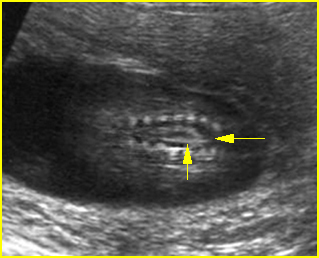
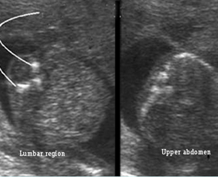
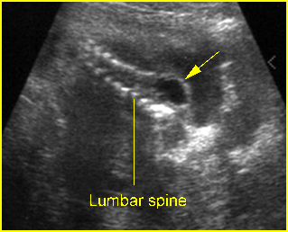
Direct signs  Fig 4, Fig 5, Fig 6, Fig 7, Fig 8, Fig 9

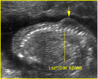
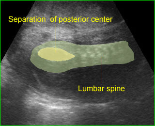
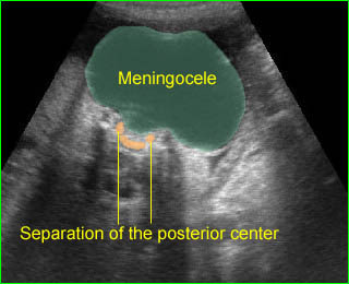
  • Visualization of the spinal dysraphic defect; widely separated ossification centers of the posterior processes, best visualized on the transverse scan (the coronal scan is also helpful). In the coronal plane, distortion of the vertebral architecture with spina bifida results in disappearance of the central line and widening of the two lines.
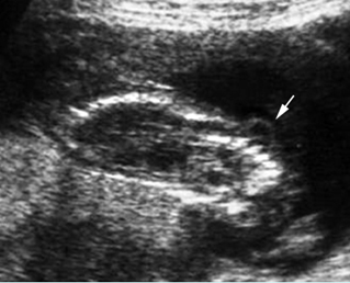
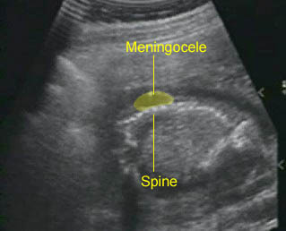
  • Demonstration of myelomeningocele sac, which is present in the majority of cases, usually covered by a thin translucent membrane and containing both CSF and neural elements, best visualized on the sagittal scan (both coronal and transverse scans are also helpful). In the sagittal scans, the posterior line and overlying soft tissues are absent at the level of the lesion and these scans are also useful for evaluating the severity of the lesion as well as spinal curvatures that may be exaggerated with large defects.

Fig 4:  Spina bifida   Coronal scan of the lumbosacral spine: separation of the ossification centers of the spine (arrow)

Fig 5:  Spina bifida  Cross-sectional scan of the lumbar and thoracic spine: separation of posterior ossification centers at the lumbar region with small protrusion of the overlying tissue

Fig 6:  Lumbar meningocele   Oblique sagittal scan of the spine: small cystic mass (arrow) protruding from the lumbar region

Fig 7:  Lumbar meningocele   Oblique sagittal scan of the spine: small protrusion (arrow) of the overlying skin at the lumbar region

Fig 8:  Lumbar meningocele   Oblique sagittal scan of the spine: small cystic mass (arrow) protruding from the lumbar region

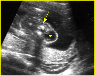
Fig 9:  Cervical meningocele  Cross-sectional scan of the cervical spine: small cystic mass (*) arising from cervical spine (arrow)

Video clips of spina bifida

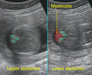
Spina bifida   Cross-sectional scan of the normal spine at the level of upper abdomen, but wide separation of the posterior ossification centers at the lumbar spine with the protrusion of the spinal cord

Spina bifida :  Coronal scan: mild separation of the posterior centers of the lumbosacral spine

Spina bifida :  Sagittal scan: small meningocele at the lumbar spine

Large meningocele :  Cross-sectional scan of the normal spine at the level of upper abdomen, but wide separation of the posterior ossification centers at the lumbar spine with the protrusion of the meningeal sac

Spina bifida :  Sagittal scan of the fetal spine showing sacral meningocele

Indirect signs

Fig 10, Fig 11

  • Lemon sign: a concave deformity or scalloping of the frontal bones at the level of the coronal suture which is best elicited on axial scans at the level of lateral ventricles. The lemon sign is a very sensitive sign for predicting spina bifida before 24 weeks with a sensitivity of 98%, but it has a low sensitivity (13%) after 24 weeks. It is not specific and is found in 1-2% of normal fetuses.
  • Cerebellar sign: obliteration of the cisterna magna caused by the caudal displacement of the cranial contents resulting in either an absent cerebellum or a flattened, centrally curved, banana-like cerebellum (banana sign). Cerebellar signs are highly sensitive (95%) irrespective of gestation; however, the sign at <24 weeks of gestation is predominantly the banana sign (72%), whereas at gestations >24 weeks it is cerebellar “absence” (81%). The effacement of the cisterna magna has a high negative predictive value and is useful during routine screening of the fetal neural axis.
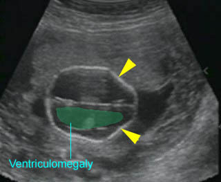
  • Ventriculomegaly: about 80% of fetuses with spina bifida demonstrate ventriculomegaly by 24 weeks.Most fetuses with open spina bifida develop ventriculomegaly, and the majority do so by 21 weeks of gestation.
  • Talipes develops after 20 weeks in most cases.
  • The sensitivity of the cranial signs is approximately 99% with a false-positive rate of 0-3%.

Fig 10:  Banana sign  Transcerebellar view: banana shape (*) of cerebellum, instead of dumbbell shape, occurring in lumbar meningocele

Fig 11:  Lemon sign  Transverse scan at the level of lateral ventricles: indent frontal bones in association with lumbar meningocele

Video clips of spina bifida

Lemon sign :  Indentation of the frontal bones (arrowhead) with ventriculomegaly

–     The accuracy of the diagnosis depends on the experience of the operator, and the quality of the equipment varies from 70% to nearly 100%. However, the sensitivity among routine non-targeted examinations is probably about 75-80%. With an experienced operator, sonography alone has a sensitivity of 97% and a specificity of 100%.

–     Normal ultrasound is an appropriate basis for a reduction of at least 95% in the maternal serum alpha-fetoprotein-based risk for neural tube defects.

The main differential diagnosis includes

  • sacrococcygeal teratoma
  • conjoined twins, especially with large tumors
  • hemangioma
  • neuroectodermal cyst.

–     Usually diagnosed in the second trimester, but possibly detected at 9-10 weeks with transvaginal ultrasound.

Pitfalls:

  • Oblique views through the lumbar spine can make the gluteus muscle look like a myelomeningocele. Three-dimensional ultrasound is helpful in this case.

Associations: Spina bifida is an isolated abnormality in most cases. However, 9-17% of cases are associated with abnormal chromosomes, especially trisomy 18 and 13. Associated anomalies other than neuromuscular deformities due to the spinal defect include hydrocephalus (most common) associated with Arnold-Chiari malformation, ACC, DWM and other extracranial anomalies.

Management: Termination of pregnancy can probably be offered when diagnosed before viability. In continuing pregnancies, delivery can occur at term. Preterm delivery may be considered in cases of rapid development of ventriculomegaly. Prelabor cesarean section may be considered to reduce motor dysfunction. Although several reports have indicated that in utero repairs of myelomeningocele decrease hindbrain herniation and shunt-dependent hydrocephalus, the operation seems to be associated with an increased rate of premature delivery, no improvement in the degree of paralysis and bladder function, and possibly pulmonary hypoplasia. The procedures should be considered as investigational.
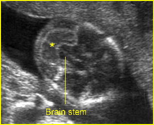
Prognosis: Depends on the size, location and the presence of ventriculomegaly, the extent of the lesion, and the presence or absence of neural tissue. Although modern surgical and medical management has resulted in better long-term function, the mortality rate and urinary tract and orthopedic disability are sill high.

Recurrence risk: Isolated neural tube defects, a multifactorial disorder, carry a recurrence risk of about 2-4% after one affected pregnancy, and 10-15% after two affected pregnancies. Folic acid periconceptional supplementation can effectively reduce the recurrent risk. 50-70% of these defects can be prevented if a woman consumes sufficient folic acid daily before conception and throughout the first trimester of her pregnancy.

Read More

Hypoplastic Thorax

Hypoplastic Thorax

The specific group of autosomal recessive disorders mainly involves ribs and thoracic hypoplasia including asphyxiating thoracic dysplasia, Ellis-van Creveld syndrome, and short-rib polydactyly syndrome. There is phenotypic overlap between these three entities. They may be a spectrum of the same entity.

Sonographic differential diagnoses of the hypoplastic thorax include:

  • Asphyxiating thoracic dysplasia (Jeune syndrome)
  • Ellis-van Creveld syndrome
  • Short-rib polydactyly syndrome
  • Other syndromes with a lesser degree of rib shortenings
    • thanatophoric dysplasia
    • atelosteogenesis
    • fibrochondrogenesis
    • achondrogenesis
    • Jarcho-Levin syndrome.

 Asphyxiating thoracic dysplasia (Jeune syndrome)

Asphyxiating thoracic dysplasia (Jeune syndrome) is an autosomal recessive skeletal dysplasia, an ATD gene located on chromosome 15q13, characterized by a small thorax, a varying degree of short limbs, renal anomaly, and polydactyly.

Incidence: Rare.

Sonographic findings:

 Fig 1, Fig 2

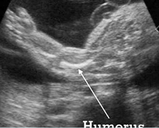
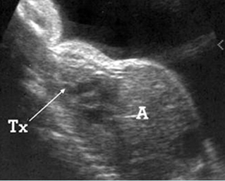
  • Narrow and bell-shaped thorax, with short, horizontal ribs.
  • Normal or mildly shortened long bones, but not as short as those in short-rib polydactyly syndrome or thanatophoric dysplasia.
  • Polydactyly and cleft lip/palate in many cases.
  • Associated renal abnormalities.
  • Normal bone echogenicity.
  •  Increased nuchal translucency at late first trimester.
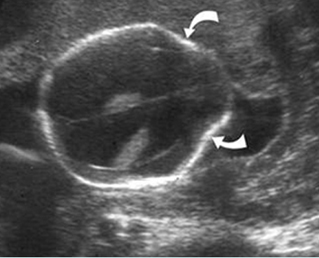
  • Associated anomalies: renal cystic dysplasia, pancreatic cyst.
  • Usually diagnosed in the second or third trimester, but diagnosis in the first trimester has been reported.
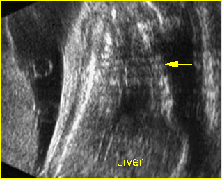
Fig 1:  Short humerus  Longitudinal scan of long bones: shortened but well ossified humerus of the fetus with Juene syndrome
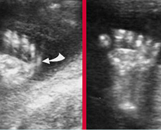
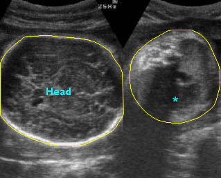
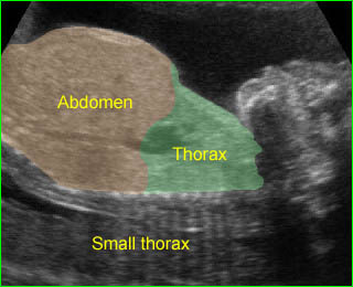
Fig 2:  Small thorax  Sagittal scan of the thorax and abdomen: disproportion in size of the thorax and abdomen of the fetus with Juene syndrome (Tx = chest, A = abdomen)

Management: Termination of pregnancy can be offered when diagnosed before viability.

Prognosis: Poor, but with a wide spectrum of severity from lethal to long-term survival.

Recurrence risk: Theoretically, the recurrent risk of these autosomal recessive disorders is 25%.

Short-rib polydactyly syndrome (SRP)

SRP is an autosomal recessive disorder characterized by short ribs, micromelia and polydactyly. SRP may be associated with a gene defect involving 4q13 or 4p16.

Incidence: Rare.

Sonographic findings:

  • Micromelia, usually severe.
  • Constricted thorax with severe short ribs.
  • Postaxial polydactyly.
  • Normal bone echogenicity.
  • Associated, genitourinary and gastrointestinal anomalies (Saldino-Noonan type).
  • 3D ultrasound may provide additional details.
  • SRP may be divided into three subtypes as follows (these subtypes are probably part of a continuous spectrum with variable expressivity):
    • associated cleft lip and palate (Majewski type)
    • associated renal abnormality (Naumoff type)
    •  associated cleft lip/palate, genitourinary, gastrointestinal anomalies (Beemer-Langer type).
  • Usually diagnosed in the second half of pregnancy but possible as early as 13 weeks.

 Management: Termination of pregnancy can be offered when diagnosed before viability.

Prognosis: Poor, but with a wide spectrum of severity from lethal to long-term survival.

Recurrence risk: Theoretically, the recurrent risk of these autosomal recessive disorders is 25%.

 Chondroectodermal Dysplasia (Ellis-van Creveld syndrome; EVC)

EVC is an autosomal recessive disorder characterized by short ribs, short limbs, polydactyly, dysplastic nails and teeth. EVC may be associated with a gene defect involving 4p16. DNA analysis for the first trimester diagnosis has been reported.

Incidence: Rare.

Sonographic findings:

Fig 3, Fig 4, Fig 5

  • Acromesomelia with normal spine and skull.
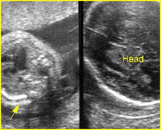
  • Long and narrow thorax with short ribs.
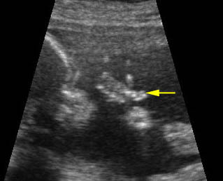
  • Postaxial polydactyly.
  • Congenital heart defects in 60% of cases.
  • Normal bone echogenicity.
  • Usually diagnosed in the second half of pregnancy but possible to diagnose as early as the late first trimester.

 Management: Termination of pregnancy may be considered when diagnosed before viability. For the continuing pregnancy, ECV should not alter the standard obstetric management.

Prognosis: Depends on the severity, which varies from lethal to long-term survival. Overall the prognosis is rather good, but there is a significant mortality rate, due primarily to cardiorespiratory failure.

Recurrence risk: Theoretically, the recurrent risk of these autosomal recessive disorders is 25%.

Fig 3:  Small thorax   Cross-sectional scan of the thorax and skull: disproportion in size of the thorax and head of the fetus with Ellis-van Creveld syndrome (arrow = short rib)

Fig 4:  Small thorax  Coronal scan: showing disproportion in size between thorax and abdomen of the fetus with Ellis-van Creveld syndrome

Fig 3:  Polydactyly

Video clips of hypoplastic thorax

Lung hypoplasia / SRP Syndrome :  Small thorax (*) with short ribs compared to the head size in case of fetal short-rib polydactyly syndrome

Post-axial polydactyly

Short-rib polydactyly syndrome :  Markedly small thorax compared to the abdomen in case of short-rib polydactyly syndrome

Read More

Diastrophic Dysplasia

Diastrophic Dysplasia

Diastrophic dysplasia, an autosomal recessive disorder related to a mutation in the DTDST gene, is characterized by micromelia, clubfoot hand deformities, multiple joint flexion contractures, and scoliosis. DNA analysis for prenatal diagnosis is possible.

Incidence: Rare.

Sonographic findings:

Fig 1, Fig 2, Fig 3, Fig 4

  • Micromelia, typically severe rhizomelic-type limb shortening and bowing of all long bones but a wide spectrum of severity.
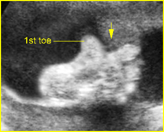
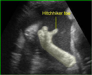
  • Hand deformities with abducted positions of the thumbs or toes called hitchhiker thumbs and toes which are relatively unique for diastrophic dysplasia; 3D ultrasound may provide better details.
  • Long bones are not bowed, unlike most other lethal syndromes.
  • Micrognathia, facial clefts and cardiac defects are occasionally seen.
  • Differential diagnoses include thanatophoric dysplasia, achondroplasia, arthrogryposis multiple congenita, atelosteosgenesis type II and pseudodiastrophic dysplasia.
  • Usually diagnosable in the second trimester, but diagnosis in the late first trimester has been reported.

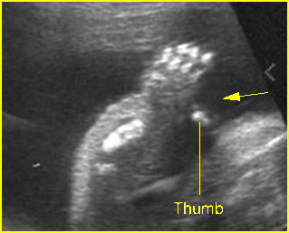
Fig 1:  Hitchhiker thumbs  Abnormal posture and wide separation of the thumb (arrow) in the fetus with diastrophic dysplasia (25 weeks)

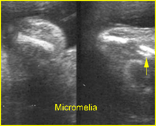
Fig 2:  Shortenings of long bones   Short humerus, ulna and radius but normal bone density in the fetus with diastrophic dysplasia

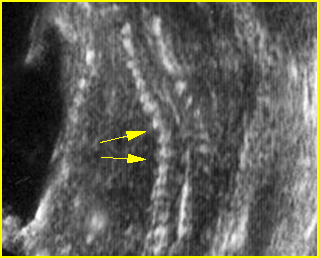
Fig 3:  Scoliosis  Coronal scan of the spine: scoliosis of thoracic spine in the fetus with diastrophic dysplasia

Fig 4:  Hitchhiker toe  Abnormal posture and wide separation of the toe (arrow) in the fetus with diastrophic dysplasia (26 weeks)

Video clips of diastrophic dysplasia

Hitchhiker toe  :  Persistent wide separation of the thumb and the second toe

Associations: Rare.

Management: Termination of pregnancy may be an option when diagnosed before viability. For the continuing pregnancy, diastrophic dysplasia should not alter the standard obstetric management.

Recurrence risk: Because it is an autosomal recessive disorder, the theoretical risk of recurrence is 25%.

Prognosis: Non-lethal, normal intellectual development, but a wide spectrum of morbidity.

Read More

Campomelic Dysplasia

Campomelic Dysplasia

Campomelic dysplasia is a rare lethal disorder characterized by bowing of long bones of the lower extremities, an enlarged and elongated skull with a peculiar small facies, and hypoplastic scapulae. This disorder is related to a single mutation in the SOX-9 gene located at 17q24, an SRY-related gene. An apparent preponderance of females with this condition is due to 46,XY male with sex reversal.

Incidence: Approximately 1 in 110,000 births.

Sonographic findings:

Fig 1, Fig 2, Fig 3

  • Bowing, sometimes angulation, of the femur and tibia.
  • Hypoplastic or absent fibulae is commonly seen.
  • Other tubular bones are normal in length.
  • Bell-shaped narrow thorax.
  • Hypoplastic scapulae.
  • Normal bone echogenicity.
  • Occasional associated deformities: clubfeet, micrognathia, pyelectasis, and ventriculomegaly.
  • 3D ultrasound may provide information supplementary to that provided by 2D ultrasound for early diagnosis.
  • The main differential diagnoses include any cause with bowing long bones such as thanatophoric dysplasia, and variants of osteogenesis imperfecta.
  • Pitfalls: A variant of the same entity disorder may show no bowed limbs, a so-called acampomelic campomelic dysplasia.
  • Usually diagnosed in the second half of pregnancy.

Fig 1:  Campomelic dysplasia   Longitudinal scan of tibia: anterior bowing of well ossified tibia

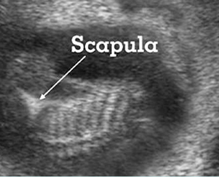
Fig 2:  Hypoplastic scapula  Coronal scan of the scapula: scapula hypoplasia of the fetus with campomelic syndrome

Fig 3:  Small thorax  Coronal scan of fetal trunk: disproportion of thorax (arrow) and abdomen size of the fetus with campomelic dysplasia

Video clips of campomelic dysplasia

Campomelia :  Anterior bowing of the tibia o (arrow)

Campomelia :  Coronal scan of the trunk:: markedly small thorax (solid circle)

Associations: Rare, several non-specific anomalies such as micrognathia, hydronephrosis and cardiac defects.

Management: Termination of pregnancy can be offered.

Prognosis: Lethal in most cases.

Recurrence risk: The majority of cases are caused by new mutations, therefore, the recurrence risk is rare.

Read More